


 Foundational characteristics of cancer include proliferation, angiogenesis, migration, evasion of apoptosis, and cellular immortality. Find key markers for these cellular processes and antibodies to detect them.
 The SUMOplot™ Analysis Program predicts and scores sumoylation sites in your protein. SUMOylation is a post-translational modification involved in various cellular processes, such as nuclear-cytosolic transport, transcriptional regulation, apoptosis, protein stability, response to stress, and progression through the cell cycle.
 The Autophagy Receptor Motif Plotter predicts and scores autophagy receptor binding sites in your protein. Identifying proteins connected to this pathway is critical to understanding the role of autophagy in physiological as well as pathological processes such as development, differentiation, neurodegenerative diseases, stress, infection, and cancer.
BRD3 Antibody - middle region
Rabbit Polyclonal Antibody
- SPECIFICATION
- CITATIONS
- PROTOCOLS
- BACKGROUND

Application
| WB |
|---|---|
| Primary Accession | Q15059 |
| Other Accession | Q15059-2 |
| Reactivity | Human |
| Predicted | Human, Mouse, Rat, Rabbit, Zebrafish, Pig, Dog, Guinea Pig, Horse, Bovine |
| Host | Rabbit |
| Clonality | Polyclonal |
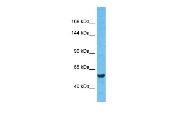
| Calculated MW | 61 kDa |
| Gene ID | 8019 |
|---|---|
| Other Names | Bromodomain-containing protein 3, RING3-like protein, BRD3, KIAA0043, RING3L |
| Target/Specificity | This gene was identified based on its homology to the gene encoding the RING3 protein, a serine/threonine kinase. The gene localizes to 9q34, a region which contains several major histocompatibility complex (MHC) genes. The function of the encoded protein is not known. |
| Format | Liquid. Purified antibody supplied in 1x PBS buffer with 0.09% (w/v) sodium azide and 2% sucrose. |
| Reconstitution & Storage | Add 50 ul, l of distilled water. Final Anti-BRD3 antibody concentration is 1 mg/ml in PBS buffer with 2% sucrose. For longer periods of storage, store at -20°C. Avoid repeat freeze-thaw cycles. |
| Precautions | BRD3 Antibody - middle region is for research use only and not for use in diagnostic or therapeutic procedures. |
| Name | BRD3 {ECO:0000303|PubMed:18406326, ECO:0000312|HGNC:HGNC:1104} |
|---|---|
| Function | Chromatin reader that recognizes and binds acetylated histones, thereby controlling gene expression and remodeling chromatin structures (PubMed:18406326, PubMed:22464331, PubMed:27105114, PubMed:32895492). Recruits transcription factors and coactivators to target gene sites, and activates RNA polymerase II machinery for transcriptional elongation (PubMed:29567837, PubMed:32895492). In vitro, binds acetylated lysine residues on the N-terminus of histone H2A, H2B, H3 and H4 (PubMed:18406326). Involved in endoderm differentiation via its association with long non-coding RNA (lncRNA) DIGIT: BRD3 undergoes liquid-liquid phase separation upon binding to lncRNA DIGIT, promoting binding to histone H3 acetylated at 'Lys-18' (H3K18ac) to induce endoderm gene expression (PubMed:32895492). Also binds non-histones acetylated proteins, such as GATA1 and GATA2: regulates transcription by promoting the binding of the transcription factor GATA1 to its targets (By similarity). |
| Cellular Location | Nucleus. Chromosome. Note=Detected on chromatin |
| Tissue Location | Ubiquitous.. |

Thousands of laboratories across the world have published research that depended on the performance of antibodies from Abcepta to advance their research. Check out links to articles that cite our products in major peer-reviewed journals, organized by research category.
info@abcepta.com, and receive a free "I Love Antibodies" mug.

Provided below are standard protocols that you may find useful for product applications.
Background
This is a rabbit polyclonal antibody against BRD3. It was validated on Western Blot by Abgent. At Abgent we manufacture rabbit polyclonal antibodies on a large scale (200-1000 products/month) of high throughput manner. Our antibodies are peptide based and protein family oriented. We usually provide antibodies covering each member of a whole protein family of your interest. We also use our best efforts to provide you antibodies recognize various epitopes of a target protein. For availability of antibody needed for your experiment, please inquire (sales@abgent.com).
If you have used an Abcepta product and would like to share how it has performed, please click on the "Submit Review" button and provide the requested information. Our staff will examine and post your review and contact you if needed.
If you have any additional inquiries please email technical services at tech@abcepta.com.
Ordering Information
Other Products
Shipping Information
