


 Foundational characteristics of cancer include proliferation, angiogenesis, migration, evasion of apoptosis, and cellular immortality. Find key markers for these cellular processes and antibodies to detect them.
 The SUMOplot™ Analysis Program predicts and scores sumoylation sites in your protein. SUMOylation is a post-translational modification involved in various cellular processes, such as nuclear-cytosolic transport, transcriptional regulation, apoptosis, protein stability, response to stress, and progression through the cell cycle.
 The Autophagy Receptor Motif Plotter predicts and scores autophagy receptor binding sites in your protein. Identifying proteins connected to this pathway is critical to understanding the role of autophagy in physiological as well as pathological processes such as development, differentiation, neurodegenerative diseases, stress, infection, and cancer.
> home > Products > Primary Antibodies > Antibody Collections > SARS CoV2 PPIs > JAKMIP1 antibody - middle region
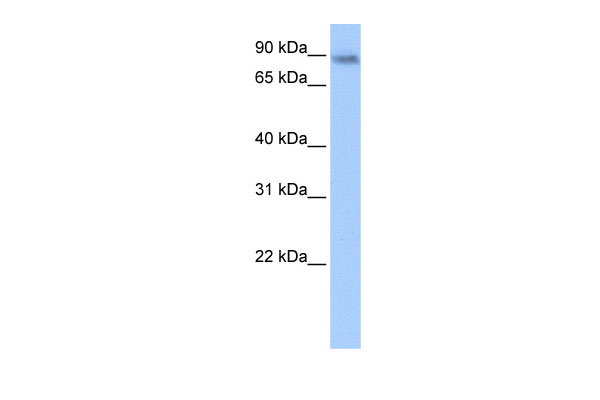
JAKMIP1 antibody - middle region
Rabbit Polyclonal Antibody
- SPECIFICATION
- CITATIONS
- PROTOCOLS
- BACKGROUND

Application
| WB |
|---|---|
| Primary Accession | Q96N16 |
| Other Accession | XM_001129178, XP_001129178 |
| Reactivity | Human, Mouse, Rat, Pig, Horse, Bovine, Dog |
| Predicted | Human, Mouse, Pig, Horse, Dog |
| Host | Rabbit |
| Clonality | Polyclonal |
| Calculated MW | 97kDa |
| Gene ID | 152789 |
|---|---|
| Alias Symbol | FLJ31564, Gababrbp, JAMIP1, MARLIN1 |
| Other Names | Janus kinase and microtubule-interacting protein 1, GABA-B receptor-binding protein, Multiple alpha-helices and RNA-linker protein 1, Marlin-1, JAKMIP1, GABABRBP, JAMIP1, MARLIN1 |
| Format | Liquid. Purified antibody supplied in 1x PBS buffer with 0.09% (w/v) sodium azide and 2% sucrose. |
| Reconstitution & Storage | Add 50 ul of distilled water. Final anti-JAKMIP1 antibody concentration is 1 mg/ml in PBS buffer with 2% sucrose. For longer periods of storage, store at 20°C. Avoid repeat freeze-thaw cycles. |
| Precautions | JAKMIP1 antibody - middle region is for research use only and not for use in diagnostic or therapeutic procedures. |
| Name | JAKMIP1 |
|---|---|
| Synonyms | GABABRBP, JAMIP1, MARLIN1 |
| Function | Associates with microtubules and may play a role in the microtubule-dependent transport of the GABA-B receptor. May play a role in JAK1 signaling and regulate microtubule cytoskeleton rearrangements. |
| Cellular Location | Cytoplasm, cytoskeleton. Membrane; Peripheral membrane protein. Note=Colocalizes with the microtubule network Localizes to the cell body and neurites of hippocampal neurons where it accumulates in granules. Localizes to the tail and to a lower extent to the head of sperm cells |
| Tissue Location | Predominantly expressed in neural tissues and lymphoid cells (at protein level). Isoform 2, isoform 3 and isoform 4 are specifically expressed in brain and retina. Isoform 1 and isoform 5 are also detected in liver, lung and skeletal muscle. Also detected in testis and to a lower extent spleen and intestine |
Research Areas
Citations (0)

Thousands of laboratories across the world have published research that depended on the performance of antibodies from Abcepta to advance their research. Check out links to articles that cite our products in major peer-reviewed journals, organized by research category.
Submit your citation using an Abcepta antibody to
info@abcepta.com, and receive a free "I Love Antibodies" mug.
info@abcepta.com, and receive a free "I Love Antibodies" mug.

Application Protocols
Provided below are standard protocols that you may find useful for product applications.
Abcepta welcomes feedback from its customers.
If you have used an Abcepta product and would like to share how it has performed, please click on the "Submit Review" button and provide the requested information. Our staff will examine and post your review and contact you if needed.
If you have any additional inquiries please email technical services at tech@abcepta.com.
$ 194.50

⚠️ Limit: 5 vials per order for discount.
Cat# AI11847
Ordering Information
United States
AlbaniaAustraliaAustriaBelgiumBosnia & HerzegovinaBrazilBulgariaCanadaCentral AmericaChinaCroatiaCyprusCzech RepublicDenmarkEstoniaFinlandFranceGermanyGreeceHong KongHungaryIcelandIndiaIndonesiaIrelandIsraelItalyJapanLatviaLithuaniaLuxembourgMacedoniaMalaysiaMaltaMexicoNetherlandsNew ZealandNorwayPakistanPolandPortugalRomaniaSerbiaSingaporeSlovakiaSloveniaSouth AfricaSouth KoreaSpainSwedenSwitzerlandTaiwanTurkeyUnited KingdomUnited StatesVietnamWorldwideOthers
USA Headquarters
(888) 735-7227 / (858) 622-0099 or (858) 875-1900
Other Products
Shipping Information
Domestic orders (in stock items)
Shipped out the same day. Orders placed after 1 PM (PST) will ship out the next business day.
International orders
Contact your local distributors
