


 Foundational characteristics of cancer include proliferation, angiogenesis, migration, evasion of apoptosis, and cellular immortality. Find key markers for these cellular processes and antibodies to detect them.
 The SUMOplot™ Analysis Program predicts and scores sumoylation sites in your protein. SUMOylation is a post-translational modification involved in various cellular processes, such as nuclear-cytosolic transport, transcriptional regulation, apoptosis, protein stability, response to stress, and progression through the cell cycle.
 The Autophagy Receptor Motif Plotter predicts and scores autophagy receptor binding sites in your protein. Identifying proteins connected to this pathway is critical to understanding the role of autophagy in physiological as well as pathological processes such as development, differentiation, neurodegenerative diseases, stress, infection, and cancer.
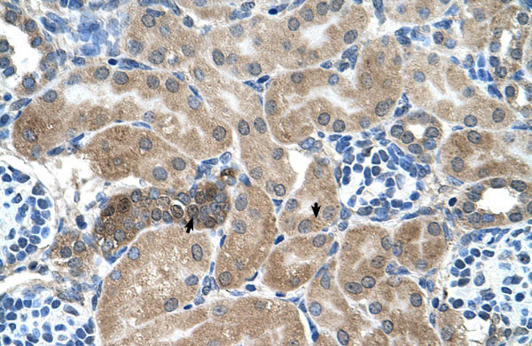
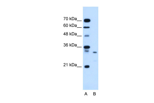
SDCBP antibody - middle region
Rabbit Polyclonal Antibody
- SPECIFICATION
- CITATIONS
- PROTOCOLS
- BACKGROUND

Application
| WB, IHC |
|---|---|
| Primary Accession | O00560 |
| Other Accession | NM_001007067, NP_001007068 |
| Reactivity | Human, Mouse, Rat, Rabbit, Zebrafish, Pig, Horse, Bovine, Guinea Pig, Dog |
| Predicted | Human, Mouse, Rabbit, Zebrafish, Pig, Chicken, Horse, Dog |
| Host | Rabbit |
| Clonality | Polyclonal |
| Calculated MW | 32kDa |
| Gene ID | 6386 |
|---|---|
| Alias Symbol | MDA-9, ST1, SYCL, TACIP18 |
| Other Names | Syntenin-1, Melanoma differentiation-associated protein 9, MDA-9, Pro-TGF-alpha cytoplasmic domain-interacting protein 18, TACIP18, Scaffold protein Pbp1, Syndecan-binding protein 1, SDCBP, MDA9, SYCL |
| Format | Liquid. Purified antibody supplied in 1x PBS buffer with 0.09% (w/v) sodium azide and 2% sucrose. |
| Reconstitution & Storage | Add 100 ul of distilled water. Final anti-SDCBP antibody concentration is 1 mg/ml in PBS buffer with 2% sucrose. For longer periods of storage, store at 20°C. Avoid repeat freeze-thaw cycles. |
| Precautions | SDCBP antibody - middle region is for research use only and not for use in diagnostic or therapeutic procedures. |
| Name | SDCBP |
|---|---|
| Synonyms | MDA9, SYCL |
| Function | Multifunctional adapter protein involved in diverse array of functions including trafficking of transmembrane proteins, neuro and immunomodulation, exosome biogenesis, and tumorigenesis (PubMed:26291527). Positively regulates TGFB1-mediated SMAD2/3 activation and TGFB1-induced epithelial-to-mesenchymal transition (EMT) and cell migration in various cell types. May increase TGFB1 signaling by enhancing cell-surface expression of TGFR1 by preventing the interaction between TGFR1 and CAV1 and subsequent CAV1-dependent internalization and degradation of TGFR1 (PubMed:25893292). In concert with SDC1/4 and PDCD6IP, regulates exosome biogenesis (PubMed:22660413). Regulates migration, growth, proliferation, and cell cycle progression in a variety of cancer types (PubMed:26539120). In adherens junctions may function to couple syndecans to cytoskeletal proteins or signaling components. Seems to couple transcription factor SOX4 to the IL-5 receptor (IL5RA) (PubMed:11498591). May also play a role in vesicular trafficking (PubMed:11179419). Seems to be required for the targeting of TGFA to the cell surface in the early secretory pathway (PubMed:10230395). |
| Cellular Location | Cell junction, focal adhesion. Cell junction, adherens junction. Cell membrane; Peripheral membrane protein. Endoplasmic reticulum membrane; Peripheral membrane protein. Nucleus. Melanosome. Cytoplasm, cytosol. Cytoplasm, cytoskeleton. Secreted, extracellular exosome. Membrane raft. Note=Mainly membrane-associated Localized to adherens junctions, focal adhesions and endoplasmic reticulum. Colocalized with actin stress fibers. Also found in the nucleus. Identified by mass spectrometry in melanosome fractions from stage I to stage IV. Associated to the plasma membrane in the presence of FZD7 and phosphatidylinositol 4,5-bisphosphate (PIP2) (PubMed:27386966). |
| Tissue Location | Expressed in lung cancers, including adenocarcinoma, squamous cell carcinoma and small-cell carcinoma (at protein level) (PubMed:25893292). Widely expressed. Expressed in fetal kidney, liver, lung and brain. In adult highest expression in heart and placenta. |

Thousands of laboratories across the world have published research that depended on the performance of antibodies from Abcepta to advance their research. Check out links to articles that cite our products in major peer-reviewed journals, organized by research category.
info@abcepta.com, and receive a free "I Love Antibodies" mug.

Provided below are standard protocols that you may find useful for product applications.
References
Boukerche,H.,(2005)CancerRes.65(23),10901-10911ReconstitutionandStorage:Forshorttermuse,storeat2-8Cupto1week.Forlongtermstorage,storeat-20Cinsmallaliquotstopreventfreeze-thawcycles.
If you have used an Abcepta product and would like to share how it has performed, please click on the "Submit Review" button and provide the requested information. Our staff will examine and post your review and contact you if needed.
If you have any additional inquiries please email technical services at tech@abcepta.com.
Ordering Information
Other Products
Shipping Information
