


 Foundational characteristics of cancer include proliferation, angiogenesis, migration, evasion of apoptosis, and cellular immortality. Find key markers for these cellular processes and antibodies to detect them.
 The SUMOplot™ Analysis Program predicts and scores sumoylation sites in your protein. SUMOylation is a post-translational modification involved in various cellular processes, such as nuclear-cytosolic transport, transcriptional regulation, apoptosis, protein stability, response to stress, and progression through the cell cycle.
 The Autophagy Receptor Motif Plotter predicts and scores autophagy receptor binding sites in your protein. Identifying proteins connected to this pathway is critical to understanding the role of autophagy in physiological as well as pathological processes such as development, differentiation, neurodegenerative diseases, stress, infection, and cancer.
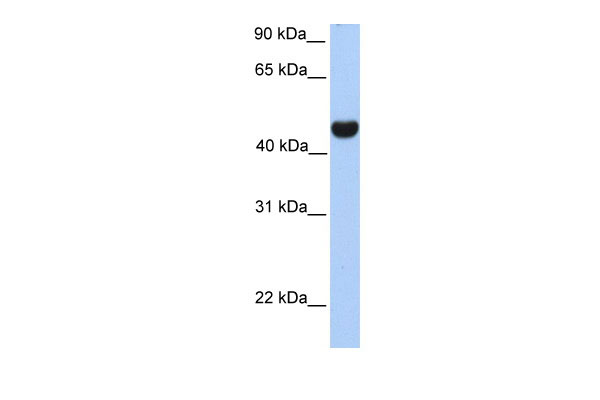
RTN4 antibody - middle region
Rabbit Polyclonal Antibody
- SPECIFICATION
- CITATIONS
- PROTOCOLS
- BACKGROUND

Application
| WB |
|---|---|
| Primary Accession | Q9NQC3 |
| Other Accession | NM_207520, NP_997403 |
| Reactivity | Human, Mouse, Rat, Rabbit, Zebrafish, Pig, Sheep, Horse, Bovine, Guinea Pig, Dog |
| Predicted | Human, Mouse, Rat, Rabbit, Pig, Chicken, Sheep, Horse, Bovine, Guinea Pig, Dog |
| Host | Rabbit |
| Clonality | Polyclonal |
| Calculated MW | 42kDa |
| Gene ID | 57142 |
|---|---|
| Alias Symbol | ASY, NI220/250, NOGO, NOGO-A, NOGOC, NSP, NSP-CL, Nbla00271, Nbla10545, Nogo-B, Nogo-C, RTN-X, RTN4-A, RTN4-B1, RTN4-B2, RTN4-C |
| Other Names | Reticulon-4, Foocen, Neurite outgrowth inhibitor, Nogo protein, Neuroendocrine-specific protein, NSP, Neuroendocrine-specific protein C homolog, RTN-x, Reticulon-5, RTN4, KIAA0886, NOGO |
| Format | Liquid. Purified antibody supplied in 1x PBS buffer with 0.09% (w/v) sodium azide and 2% sucrose. |
| Reconstitution & Storage | Add 50 ul of distilled water. Final anti-RTN4 antibody concentration is 1 mg/ml in PBS buffer with 2% sucrose. For longer periods of storage, store at 20°C. Avoid repeat freeze-thaw cycles. |
| Precautions | RTN4 antibody - middle region is for research use only and not for use in diagnostic or therapeutic procedures. |
| Name | RTN4 (HGNC:14085) |
|---|---|
| Function | Required to induce the formation and stabilization of endoplasmic reticulum (ER) tubules (PubMed:24262037, PubMed:25612671, PubMed:27619977). They regulate membrane morphogenesis in the ER by promoting tubular ER production (PubMed:24262037, PubMed:25612671, PubMed:27619977, PubMed:27786289). They influence nuclear envelope expansion, nuclear pore complex formation and proper localization of inner nuclear membrane proteins (PubMed:26906412). However each isoform have specific functions mainly depending on their tissue expression specificities (Probable). |
| Cellular Location | [Isoform A]: Endoplasmic reticulum membrane; Multi-pass membrane protein. Cell membrane; Multi-pass membrane protein; Cytoplasmic side Synapse {ECO:0000250|UniProtKB:Q99P72}. Note=Anchored to the membrane of the endoplasmic reticulum (ER) through 2 putative transmembrane domains. Localizes throughout the ER tubular network (PubMed:27619977) Co-localizes with TMEM33 at the ER sheets [Isoform C]: Endoplasmic reticulum membrane; Multi-pass membrane protein |
| Tissue Location | Isoform A: is specifically expressed in brain and testis and weakly in heart and skeletal muscle. Isoform B: widely expressed except for the liver. Highly expressed in endothelial cells and vascular smooth muscle cells, including blood vessels and mesenteric arteries (PubMed:15034570, PubMed:21183689). Isoform C: is expressed in brain, skeletal muscle and adipocytes. Isoform D is testis-specific. |

Thousands of laboratories across the world have published research that depended on the performance of antibodies from Abcepta to advance their research. Check out links to articles that cite our products in major peer-reviewed journals, organized by research category.
info@abcepta.com, and receive a free "I Love Antibodies" mug.

Provided below are standard protocols that you may find useful for product applications.
References
Hu,F.(2008)J.Neurosci.28(5),1262-1269ReconstitutionandStorage:Forshorttermuse,storeat2-8Cupto1week.Forlongtermstorage,storeat-20Cinsmallaliquotstopreventfreeze-thawcycles.Publications:Beetz,C.etal.AspasticparaplegiamousemodelrevealsREEP1-dependentERshaping.J.Clin.Invest.123,4273-82(2013).WB,Human,Bovine,Dog,Rat,Horse,Rabbit,Sheep,Pig,Mouse,Guineapig,Zebrafish24051375
If you have used an Abcepta product and would like to share how it has performed, please click on the "Submit Review" button and provide the requested information. Our staff will examine and post your review and contact you if needed.
If you have any additional inquiries please email technical services at tech@abcepta.com.
Ordering Information
Other Products
Shipping Information
