


 Foundational characteristics of cancer include proliferation, angiogenesis, migration, evasion of apoptosis, and cellular immortality. Find key markers for these cellular processes and antibodies to detect them.
 The SUMOplot™ Analysis Program predicts and scores sumoylation sites in your protein. SUMOylation is a post-translational modification involved in various cellular processes, such as nuclear-cytosolic transport, transcriptional regulation, apoptosis, protein stability, response to stress, and progression through the cell cycle.
 The Autophagy Receptor Motif Plotter predicts and scores autophagy receptor binding sites in your protein. Identifying proteins connected to this pathway is critical to understanding the role of autophagy in physiological as well as pathological processes such as development, differentiation, neurodegenerative diseases, stress, infection, and cancer.
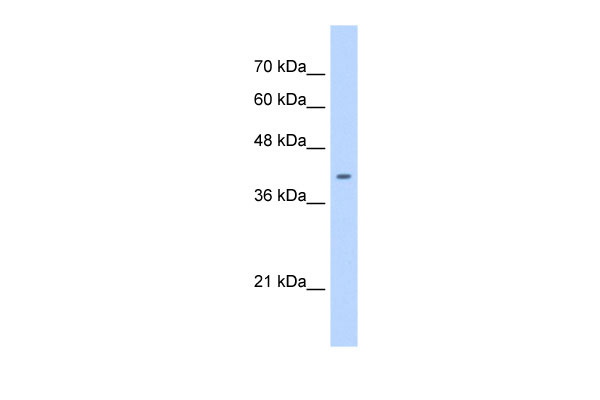
ST6GALNAC6 antibody - C-terminal region
Rabbit Polyclonal Antibody
- SPECIFICATION
- CITATIONS
- PROTOCOLS
- BACKGROUND

Application
| WB |
|---|---|
| Primary Accession | Q969X2 |
| Other Accession | NM_013443, NP_038471 |
| Reactivity | Human, Mouse, Rat, Rabbit, Pig, Horse, Bovine, Guinea Pig, Dog |
| Predicted | Human, Mouse, Rabbit, Pig, Horse, Guinea Pig, Dog |
| Host | Rabbit |
| Clonality | Polyclonal |
| Calculated MW | 38kDa |
| Gene ID | 30815 |
|---|---|
| Alias Symbol | RP11-203J24.3, SIAT7F, ST6GALNACVI |
| Other Names | Alpha-N-acetylgalactosaminide alpha-2, 6-sialyltransferase 6, 2.4.99.-, GalNAc alpha-2, 6-sialyltransferase VI, ST6GalNAc VI, ST6GalNAcVI, hST6GalNAc VI, Sialyltransferase 7F, SIAT7-F, ST6GALNAC6, SIAT7F |
| Format | Liquid. Purified antibody supplied in 1x PBS buffer with 0.09% (w/v) sodium azide and 2% sucrose. |
| Reconstitution & Storage | Add 50 ul of distilled water. Final anti-ST6GALNAC6 antibody concentration is 1 mg/ml in PBS buffer with 2% sucrose. For longer periods of storage, store at 20°C. Avoid repeat freeze-thaw cycles. |
| Precautions | ST6GALNAC6 antibody - C-terminal region is for research use only and not for use in diagnostic or therapeutic procedures. |
| Name | ST6GALNAC6 |
|---|---|
| Synonyms | SIAT7F |
| Function | Transfers the sialyl group (N-acetyl-alpha-neuraminyl or NeuAc) from CMP-NeuAc onto glycoproteins and glycolipids, forming an alpha-2,6-linkage. Produces branched type disialyl structures by transfer of a sialyl group onto the GalNAc or GlcNAc residue inside backbone core chains having a terminal sialic acid with an alpha-2,3- linkage on Gal. ST6GalNAcVI prefers glycolipids to glycoproteins, predominantly catalyzing the biosynthesis of ganglioside GD1alpha from GM1b (PubMed:12668675, PubMed:17123352). Besides GMb1, MSGG and other glycolipids, it shows activity towards sialyl Lc4Cer generating disialyl Lc4Cer, which can lead to the synthesis of disialyl Lewis a (Le(a)), suggested to be a cancer-associated antigen (PubMed:12668675). Also has activity toward GD1a and GT1b, and can generate DSGG (disialylgalactosylgloboside) from MSGG (monosialylgalactosylgloboside) (By similarity). |
| Cellular Location | Golgi apparatus membrane; Single- pass type II membrane protein |
| Tissue Location | Expressed in kidney, in proximal tubule epithelial cells. Expressed in colon cell lines. |

Thousands of laboratories across the world have published research that depended on the performance of antibodies from Abcepta to advance their research. Check out links to articles that cite our products in major peer-reviewed journals, organized by research category.
info@abcepta.com, and receive a free "I Love Antibodies" mug.

Provided below are standard protocols that you may find useful for product applications.
References
Senda,M.,(2007)Biochem.J.402(3),459-470ReconstitutionandStorage:Forshorttermuse,storeat2-8Cupto1week.Forlongtermstorage,storeat-20Cinsmallaliquotstopreventfreeze-thawcycles.
If you have used an Abcepta product and would like to share how it has performed, please click on the "Submit Review" button and provide the requested information. Our staff will examine and post your review and contact you if needed.
If you have any additional inquiries please email technical services at tech@abcepta.com.
Ordering Information
Other Products
Shipping Information
