


 Foundational characteristics of cancer include proliferation, angiogenesis, migration, evasion of apoptosis, and cellular immortality. Find key markers for these cellular processes and antibodies to detect them.
 The SUMOplot™ Analysis Program predicts and scores sumoylation sites in your protein. SUMOylation is a post-translational modification involved in various cellular processes, such as nuclear-cytosolic transport, transcriptional regulation, apoptosis, protein stability, response to stress, and progression through the cell cycle.
 The Autophagy Receptor Motif Plotter predicts and scores autophagy receptor binding sites in your protein. Identifying proteins connected to this pathway is critical to understanding the role of autophagy in physiological as well as pathological processes such as development, differentiation, neurodegenerative diseases, stress, infection, and cancer.
> home > Products > Primary Antibodies > Antibody Collections > Zebrafish > Mafk antibody - N-terminal region
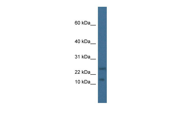
Mafk antibody - N-terminal region
Rabbit Polyclonal Antibody
- SPECIFICATION
- CITATIONS
- PROTOCOLS
- BACKGROUND

Application
| WB |
|---|---|
| Primary Accession | Q61827 |
| Other Accession | NM_010757, NP_034887 |
| Reactivity | Human, Mouse, Rat, Zebrafish, Pig, Bovine, Guinea Pig, Dog |
| Predicted | Human, Mouse, Rat, Chicken, Bovine |
| Host | Rabbit |
| Clonality | Polyclonal |
| Calculated MW | 17kDa |
| Gene ID | 17135 |
|---|---|
| Alias Symbol | AW061068, NF-E2, Nfe2u |
| Other Names | Transcription factor MafK, Erythroid transcription factor NF-E2 p18 subunit, Mafk, Nfe2u |
| Format | Liquid. Purified antibody supplied in 1x PBS buffer with 0.09% (w/v) sodium azide and 2% sucrose. |
| Reconstitution & Storage | Add 50 ul of distilled water. Final anti-Mafk antibody concentration is 1 mg/ml in PBS buffer with 2% sucrose. For longer periods of storage, store at 20°C. Avoid repeat freeze-thaw cycles. |
| Precautions | Mafk antibody - N-terminal region is for research use only and not for use in diagnostic or therapeutic procedures. |
| Name | Mafk |
|---|---|
| Synonyms | Nfe2u |
| Function | Since they lack a putative transactivation domain, the small Mafs behave as transcriptional repressors when they dimerize among themselves (By similarity). However, they act as transcriptional activators by dimerizing with other (usually larger) basic-zipper proteins, such as NFE2, NFE2L1/NRF1, NFE2L2/NRF2 and NFE2L3/NRF3, and recruiting them to specific DNA-binding sites (PubMed:9240432). Small Maf proteins heterodimerize with Fos and may act as competitive repressors of the NF-E2 transcription factor (By similarity). |
| Cellular Location | Nucleus. |
| Tissue Location | Highly expressed in heart, skeletal muscle and placenta. Also expressed in erythroid cells |
Research Areas
Citations (0)

Thousands of laboratories across the world have published research that depended on the performance of antibodies from Abcepta to advance their research. Check out links to articles that cite our products in major peer-reviewed journals, organized by research category.
Submit your citation using an Abcepta antibody to
info@abcepta.com, and receive a free "I Love Antibodies" mug.
info@abcepta.com, and receive a free "I Love Antibodies" mug.

Application Protocols
Provided below are standard protocols that you may find useful for product applications.
Abcepta welcomes feedback from its customers.
If you have used an Abcepta product and would like to share how it has performed, please click on the "Submit Review" button and provide the requested information. Our staff will examine and post your review and contact you if needed.
If you have any additional inquiries please email technical services at tech@abcepta.com.
$ 389.00
Cat# AI12810
Ordering Information
United States
AlbaniaAustraliaAustriaBelgiumBosnia & HerzegovinaBrazilBulgariaCanadaCentral AmericaChinaCroatiaCyprusCzech RepublicDenmarkEstoniaFinlandFranceGermanyGreeceHong KongHungaryIcelandIndiaIndonesiaIrelandIsraelItalyJapanLatviaLithuaniaLuxembourgMacedoniaMalaysiaMaltaMexicoNetherlandsNew ZealandNorwayPakistanPolandPortugalRomaniaSerbiaSingaporeSlovakiaSloveniaSouth AfricaSouth KoreaSpainSwedenSwitzerlandTaiwanTurkeyUnited KingdomUnited StatesVietnamWorldwideOthers
USA Headquarters
(888) 735-7227 / (858) 622-0099 or (858) 875-1900
Other Products
Shipping Information
Domestic orders (in stock items)
Shipped out the same day. Orders placed after 1 PM (PST) will ship out the next business day.
International orders
Contact your local distributors
