


 Foundational characteristics of cancer include proliferation, angiogenesis, migration, evasion of apoptosis, and cellular immortality. Find key markers for these cellular processes and antibodies to detect them.
 The SUMOplot™ Analysis Program predicts and scores sumoylation sites in your protein. SUMOylation is a post-translational modification involved in various cellular processes, such as nuclear-cytosolic transport, transcriptional regulation, apoptosis, protein stability, response to stress, and progression through the cell cycle.
 The Autophagy Receptor Motif Plotter predicts and scores autophagy receptor binding sites in your protein. Identifying proteins connected to this pathway is critical to understanding the role of autophagy in physiological as well as pathological processes such as development, differentiation, neurodegenerative diseases, stress, infection, and cancer.
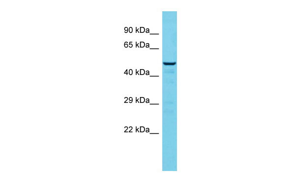
Casp2 Antibody - middle region
Rabbit Polyclonal Antibody
- SPECIFICATION
- CITATIONS
- PROTOCOLS
- BACKGROUND

Application
| WB |
|---|---|
| Primary Accession | P29594 |
| Other Accession | NM_007610, NP_031636 |
| Reactivity | Human, Mouse, Rat, Rabbit, Pig, Horse, Bovine, Guinea Pig, Dog |
| Predicted | Human, Mouse, Rat, Rabbit, Pig, Horse, Bovine, Guinea Pig, Dog |
| Host | Rabbit |
| Clonality | Polyclonal |
| Calculated MW | 51kDa |
| Gene ID | 12366 |
|---|---|
| Alias Symbol | Caspase-2, ICH-1, Nedd2 |
| Other Names | Caspase-2, CASP-2, 3.4.22.55, Neural precursor cell expressed developmentally down-regulated protein 2, NEDD-2, Protease ICH-1, Caspase-2 subunit p18, Caspase-2 subunit p13, Caspase-2 subunit p12, Casp2, Ich1, Nedd-2, Nedd2 |
| Format | Liquid. Purified antibody supplied in 1x PBS buffer with 0.09% (w/v) sodium azide and 2% sucrose. |
| Reconstitution & Storage | Add 50 ul of distilled water. Final anti-Casp2 antibody concentration is 1 mg/ml in PBS buffer with 2% sucrose. For longer periods of storage, store at 20°C. Avoid repeat freeze-thaw cycles. |
| Precautions | Casp2 Antibody - middle region is for research use only and not for use in diagnostic or therapeutic procedures. |
| Name | Casp2 |
|---|---|
| Synonyms | Ich1, Nedd-2, Nedd2 |
| Function | Involved in the activation cascade of caspases responsible for apoptosis execution. Might function by either activating some proteins required for cell death or inactivating proteins necessary for cell survival (PubMed:7958843). Associates with PIDD1 and CRADD to form the PIDDosome, a complex that activates CASP2 and triggers apoptosis in response to genotoxic stress (By similarity). |
| Tissue Location | High level expression seen in the embryonic CNS, liver, lung, kidney, small intestine, and hair follicles of vibrissae Moderate expression seen in the skin, oral mucosa, skeletal muscle, submandibular gland and thymus. In the adult, it is highly expressed in spleen, lung and kidney. Moderately in the brain, heart, testis, liver Low levels in the thymus, skeletal muscle, ovary and gut |

Thousands of laboratories across the world have published research that depended on the performance of antibodies from Abcepta to advance their research. Check out links to articles that cite our products in major peer-reviewed journals, organized by research category.
info@abcepta.com, and receive a free "I Love Antibodies" mug.

Provided below are standard protocols that you may find useful for product applications.
References
Kumar S.,et al.Genes Dev. 8:1613-1626(1994).
van de Craen M.,et al.FEBS Lett. 403:61-69(1997).
Carninci P.,et al.Science 309:1559-1563(2005).
Kumar S.,et al.Biochem. Biophys. Res. Commun. 185:1155-1161(1992).
If you have used an Abcepta product and would like to share how it has performed, please click on the "Submit Review" button and provide the requested information. Our staff will examine and post your review and contact you if needed.
If you have any additional inquiries please email technical services at tech@abcepta.com.
Ordering Information
Other Products
Shipping Information
