


 Foundational characteristics of cancer include proliferation, angiogenesis, migration, evasion of apoptosis, and cellular immortality. Find key markers for these cellular processes and antibodies to detect them.
 The SUMOplot™ Analysis Program predicts and scores sumoylation sites in your protein. SUMOylation is a post-translational modification involved in various cellular processes, such as nuclear-cytosolic transport, transcriptional regulation, apoptosis, protein stability, response to stress, and progression through the cell cycle.
 The Autophagy Receptor Motif Plotter predicts and scores autophagy receptor binding sites in your protein. Identifying proteins connected to this pathway is critical to understanding the role of autophagy in physiological as well as pathological processes such as development, differentiation, neurodegenerative diseases, stress, infection, and cancer.
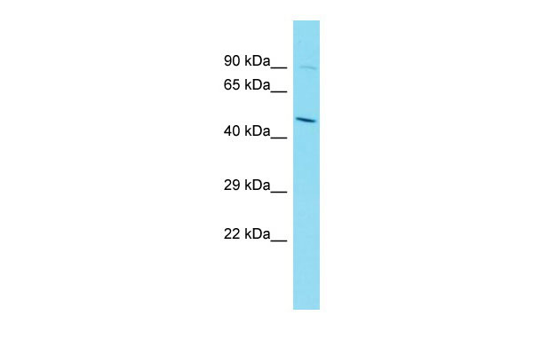
Rassf5 Antibody - C-terminal region
Rabbit Polyclonal Antibody
- SPECIFICATION
- CITATIONS
- PROTOCOLS
- BACKGROUND

Application
| WB |
|---|---|
| Primary Accession | Q5EBH1 |
| Other Accession | NM_018750, NP_061220 |
| Reactivity | Human, Mouse, Rat, Rabbit, Pig, Horse, Bovine, Guinea Pig, Dog |
| Predicted | Human, Mouse, Rat, Rabbit, Pig, Horse, Bovine, Guinea Pig, Dog |
| Host | Rabbit |
| Clonality | Polyclonal |
| Calculated MW | 47kDa |
| Gene ID | 54354 |
|---|---|
| Alias Symbol | 1300019G20Rik, AU042887, Maxp1, Nore1, Nore1A, Nore1B, Rapl |
| Other Names | Ras association domain-containing protein 5, New ras effector 1, Regulator for cell adhesion and polarization enriched in lymphoid tissues, RAPL, Rassf5, Nore1, Rapl |
| Format | Liquid. Purified antibody supplied in 1x PBS buffer with 0.09% (w/v) sodium azide and 2% sucrose. |
| Reconstitution & Storage | Add 50 ul of distilled water. Final anti-Rassf5 antibody concentration is 1 mg/ml in PBS buffer with 2% sucrose. For longer periods of storage, store at 20°C. Avoid repeat freeze-thaw cycles. |
| Precautions | Rassf5 Antibody - C-terminal region is for research use only and not for use in diagnostic or therapeutic procedures. |
| Name | Rassf5 |
|---|---|
| Synonyms | Nore1, Rapl |
| Function | Potential tumor suppressor. Seems to be involved in lymphocyte adhesion by linking RAP1A activation upon T-cell receptor or chemokine stimulation to integrin activation. Isoform 2 stimulates lymphocyte polarization and the patch-like distribution of ITGAL/LFA-1, resulting in an enhanced adhesion to ICAM1. Together with RAP1A may participate in regulation of microtubule growth. The association of isoform 2 with activated RAP1A is required for directional movement of endothelial cells during wound healing (By similarity). May be involved in regulation of Ras apoptotic function. The RASSF5-STK4/MST1 complex may mediate HRAS and KRAS induced apoptosis. |
| Cellular Location | Cytoplasm. Cytoplasm, cytoskeleton. Note=Isoform 2 is mainly located in the perinuclear region of unstimulated primary T-cells. Upon stimulation translocates to the leading edge and colocalizes with ITGAL/LFA-1 in the peripheral zone of the immunological synapse. Isoform 2 is localized to growing microtubules in vascular endothelial cells and is dissociated from microtubules by activated RAP1A (By similarity). |

Thousands of laboratories across the world have published research that depended on the performance of antibodies from Abcepta to advance their research. Check out links to articles that cite our products in major peer-reviewed journals, organized by research category.
info@abcepta.com, and receive a free "I Love Antibodies" mug.

Provided below are standard protocols that you may find useful for product applications.
References
Vavvas D.,et al.J. Biol. Chem. 273:5439-5442(1998).
Katagiri K.,et al.Nat. Immunol. 4:741-748(2003).
Okazaki N.,et al.DNA Res. 11:127-135(2004).
Carninci P.,et al.Science 309:1559-1563(2005).
Khokhlatchev A.,et al.Curr. Biol. 12:253-265(2002).
If you have used an Abcepta product and would like to share how it has performed, please click on the "Submit Review" button and provide the requested information. Our staff will examine and post your review and contact you if needed.
If you have any additional inquiries please email technical services at tech@abcepta.com.
Ordering Information
Other Products
Shipping Information
