


 Foundational characteristics of cancer include proliferation, angiogenesis, migration, evasion of apoptosis, and cellular immortality. Find key markers for these cellular processes and antibodies to detect them.
 The SUMOplot™ Analysis Program predicts and scores sumoylation sites in your protein. SUMOylation is a post-translational modification involved in various cellular processes, such as nuclear-cytosolic transport, transcriptional regulation, apoptosis, protein stability, response to stress, and progression through the cell cycle.
 The Autophagy Receptor Motif Plotter predicts and scores autophagy receptor binding sites in your protein. Identifying proteins connected to this pathway is critical to understanding the role of autophagy in physiological as well as pathological processes such as development, differentiation, neurodegenerative diseases, stress, infection, and cancer.
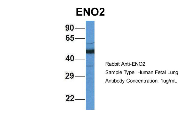
ENO2 antibody - middle region
Rabbit Polyclonal Antibody
- SPECIFICATION
- CITATIONS
- PROTOCOLS
- BACKGROUND

Application
| WB |
|---|---|
| Primary Accession | P09104 |
| Other Accession | NM_001975, NP_001966 |
| Reactivity | Human, Mouse, Rat, Rabbit, Goat, Sheep, Horse, Bovine, Guinea Pig, Dog |
| Predicted | Human, Mouse, Rabbit, Chicken, Horse, Bovine, Guinea Pig |
| Host | Rabbit |
| Clonality | Polyclonal |
| Calculated MW | 47kDa |
| Gene ID | 2026 |
|---|---|
| Alias Symbol | NSE |
| Other Names | Gamma-enolase, 4.2.1.11, 2-phospho-D-glycerate hydro-lyase, Enolase 2, Neural enolase, Neuron-specific enolase, NSE, ENO2 |
| Format | Liquid. Purified antibody supplied in 1x PBS buffer with 0.09% (w/v) sodium azide and 2% sucrose. |
| Reconstitution & Storage | Add 50 ul of distilled water. Final anti-ENO2 antibody concentration is 1 mg/ml in PBS buffer with 2% sucrose. For longer periods of storage, store at 20°C. Avoid repeat freeze-thaw cycles. |
| Precautions | ENO2 antibody - middle region is for research use only and not for use in diagnostic or therapeutic procedures. |
| Name | ENO2 |
|---|---|
| Function | Has neurotrophic and neuroprotective properties on a broad spectrum of central nervous system (CNS) neurons. Binds, in a calcium- dependent manner, to cultured neocortical neurons and promotes cell survival (By similarity). |
| Cellular Location | Cytoplasm. Cell membrane. Note=Can translocate to the plasma membrane in either the homodimeric (alpha/alpha) or heterodimeric (alpha/gamma) form |
| Tissue Location | The alpha/alpha homodimer is expressed in embryo and in most adult tissues. The alpha/beta heterodimer and the beta/beta homodimer are found in striated muscle, and the alpha/gamma heterodimer and the gamma/gamma homodimer in neurons |

Thousands of laboratories across the world have published research that depended on the performance of antibodies from Abcepta to advance their research. Check out links to articles that cite our products in major peer-reviewed journals, organized by research category.
info@abcepta.com, and receive a free "I Love Antibodies" mug.

Provided below are standard protocols that you may find useful for product applications.
References
McAleese S.M.,et al.Eur. J. Biochem. 178:413-417(1988).
van Obberghen E.,et al.J. Neurosci. Res. 19:450-456(1988).
Oliva D.,et al.Gene 79:355-360(1989).
Oliva D.,et al.Genomics 10:157-165(1991).
Ansari-Lari M.A.,et al.Genome Res. 7:268-280(1997).
If you have used an Abcepta product and would like to share how it has performed, please click on the "Submit Review" button and provide the requested information. Our staff will examine and post your review and contact you if needed.
If you have any additional inquiries please email technical services at tech@abcepta.com.
Ordering Information
Other Products
Shipping Information
