


 Foundational characteristics of cancer include proliferation, angiogenesis, migration, evasion of apoptosis, and cellular immortality. Find key markers for these cellular processes and antibodies to detect them.
 The SUMOplot™ Analysis Program predicts and scores sumoylation sites in your protein. SUMOylation is a post-translational modification involved in various cellular processes, such as nuclear-cytosolic transport, transcriptional regulation, apoptosis, protein stability, response to stress, and progression through the cell cycle.
 The Autophagy Receptor Motif Plotter predicts and scores autophagy receptor binding sites in your protein. Identifying proteins connected to this pathway is critical to understanding the role of autophagy in physiological as well as pathological processes such as development, differentiation, neurodegenerative diseases, stress, infection, and cancer.
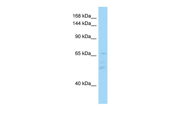
IL16 Antibody - C-terminal region
Rabbit Polyclonal Antibody
- SPECIFICATION
- CITATIONS
- PROTOCOLS
- BACKGROUND

Application
| WB |
|---|---|
| Primary Accession | Q14005 |
| Other Accession | NM_004513, NP_004504 |
| Reactivity | Human, Pig, Goat, Horse |
| Predicted | Human, Pig, Goat, Horse |
| Host | Rabbit |
| Clonality | Polyclonal |
| Calculated MW | 67kDa |
| Gene ID | 3603 |
|---|---|
| Alias Symbol | FLJ16806, FLJ42735, FLJ44234, LCF, NIL16, PRIL16, prIL-16 |
| Other Names | Pro-interleukin-16, Interleukin-16, IL-16, Lymphocyte chemoattractant factor, LCF, IL16 |
| Format | Liquid. Purified antibody supplied in 1x PBS buffer with 0.09% (w/v) sodium azide and 2% sucrose. |
| Reconstitution & Storage | Add 50 ul of distilled water. Final anti-IL16 antibody concentration is 1 mg/ml in PBS buffer with 2% sucrose. For longer periods of storage, store at 20°C. Avoid repeat freeze-thaw cycles. |
| Precautions | IL16 Antibody - C-terminal region is for research use only and not for use in diagnostic or therapeutic procedures. |
| Name | IL16 |
|---|---|
| Function | Interleukin-16 stimulates a migratory response in CD4+ lymphocytes, monocytes, and eosinophils. Primes CD4+ T-cells for IL-2 and IL-15 responsiveness. Also induces T-lymphocyte expression of interleukin 2 receptor. Ligand for CD4. Isoform 3 is involved in cell cycle progression in T-cells. Appears to be involved in transcriptional regulation of SKP2 and is probably part of a transcriptional repression complex on the core promoter of the SKP2 gene. May act as a scaffold for GABPB1 (the DNA- binding subunit the GABP transcription factor complex) and HDAC3 thus maintaining transcriptional repression and blocking cell cycle progression in resting T-cells. |
| Cellular Location | [Interleukin-16]: Secreted. [Isoform 3]: Cytoplasm. Nucleus. |
| Tissue Location | [Isoform 3]: Expressed in hemopoietic tissues, such as resting T-cells, but undetectable during active T-cell proliferation |

Thousands of laboratories across the world have published research that depended on the performance of antibodies from Abcepta to advance their research. Check out links to articles that cite our products in major peer-reviewed journals, organized by research category.
info@abcepta.com, and receive a free "I Love Antibodies" mug.

Provided below are standard protocols that you may find useful for product applications.
References
Bannert N.,et al.J. Biol. Chem. 278:42190-42199(2003).
Wang P.,et al.BMC Genomics 10:518-518(2009).
Kornfeld H.,et al.Submitted (JAN-1999) to the EMBL/GenBank/DDBJ databases.
Ota T.,et al.Nat. Genet. 36:40-45(2004).
Zody M.C.,et al.Nature 440:671-675(2006).
If you have used an Abcepta product and would like to share how it has performed, please click on the "Submit Review" button and provide the requested information. Our staff will examine and post your review and contact you if needed.
If you have any additional inquiries please email technical services at tech@abcepta.com.
Ordering Information
Other Products
Shipping Information
