


 Foundational characteristics of cancer include proliferation, angiogenesis, migration, evasion of apoptosis, and cellular immortality. Find key markers for these cellular processes and antibodies to detect them.
 The SUMOplot™ Analysis Program predicts and scores sumoylation sites in your protein. SUMOylation is a post-translational modification involved in various cellular processes, such as nuclear-cytosolic transport, transcriptional regulation, apoptosis, protein stability, response to stress, and progression through the cell cycle.
 The Autophagy Receptor Motif Plotter predicts and scores autophagy receptor binding sites in your protein. Identifying proteins connected to this pathway is critical to understanding the role of autophagy in physiological as well as pathological processes such as development, differentiation, neurodegenerative diseases, stress, infection, and cancer.
ABI2 Antibody - N-terminal region
Rabbit Polyclonal Antibody
- SPECIFICATION
- CITATIONS
- PROTOCOLS
- BACKGROUND

Application
| WB |
|---|---|
| Primary Accession | Q9NYB9 |
| Other Accession | NM_005759, NP_005750 |
| Reactivity | Human, Mouse, Rat, Rabbit, Horse, Yeast, Bovine, Guinea Pig, Dog |
| Predicted | Human, Mouse, Rat, Rabbit, Pig, Horse, Yeast, Bovine, Guinea Pig, Dog |
| Host | Rabbit |
| Clonality | Polyclonal |
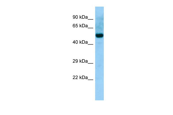
| Calculated MW | 52kDa |
| Gene ID | 10152 |
|---|---|
| Alias Symbol | ABI-2, ABI2B, AIP-1, AblBP3, SSH3BP2, argBPIA, argBPIB |
| Other Names | Abl interactor 2, Abelson interactor 2, Abi-2, Abl-binding protein 3, AblBP3, Arg-binding protein 1, ArgBP1, ABI2, ARGBPIA |
| Format | Liquid. Purified antibody supplied in 1x PBS buffer with 0.09% (w/v) sodium azide and 2% sucrose. |
| Reconstitution & Storage | Add 50 ul of distilled water. Final anti-ABI2 antibody concentration is 1 mg/ml in PBS buffer with 2% sucrose. For longer periods of storage, store at 20°C. Avoid repeat freeze-thaw cycles. |
| Precautions | ABI2 Antibody - N-terminal region is for research use only and not for use in diagnostic or therapeutic procedures. |
| Name | ABI2 {ECO:0000303|PubMed:28397838, ECO:0000312|HGNC:HGNC:24011} |
|---|---|
| Function | Regulator of actin cytoskeleton dynamics underlying cell motility and adhesion. Functions as a component of the WAVE complex, which activates actin nucleating machinery Arp2/3 to drive lamellipodia formation (PubMed:21107423). Acts as a regulator and substrate of nonreceptor tyrosine kinases ABL1 and ABL2 involved in processes linked to cell growth and differentiation. Positively regulates ABL1-mediated phosphorylation of ENAH, which is required for proper polymerization of nucleated actin filaments at the leading edge (PubMed:10498863, PubMed:7590236, PubMed:8649853). Contributes to the regulation of actin assembly at the tips of neuron projections. In particular, controls dendritic spine morphogenesis and may promote dendritic spine specification toward large mushroom-type spines known as repositories of memory in the brain (By similarity). In hippocampal neurons, may mediate actin-dependent BDNF-NTRK2 early endocytic trafficking that triggers dendrite outgrowth (By similarity). Participates in ocular lens morphogenesis, likely by regulating lamellipodia-driven adherens junction formation at the epithelial cell-secondary lens fiber interface (By similarity). Also required for nascent adherens junction assembly in epithelial cells (PubMed:15572692). |
| Cellular Location | Cytoplasm. Nucleus |
| Tissue Location | Widely expressed. Abundant in testes, ovary, thymus, and colon, with lower but detectable levels in prostate, peripheral blood leukocytes, and spleen. |

Thousands of laboratories across the world have published research that depended on the performance of antibodies from Abcepta to advance their research. Check out links to articles that cite our products in major peer-reviewed journals, organized by research category.
info@abcepta.com, and receive a free "I Love Antibodies" mug.

Provided below are standard protocols that you may find useful for product applications.
References
Dai Z.,et al.Genes Dev. 9:2569-2582(1995).
Ren R.,et al.Submitted (JUL-1995) to the EMBL/GenBank/DDBJ databases.
Wang B.,et al.Oncogene 12:1921-1929(1996).
Kalnine N.,et al.Submitted (AUG-2003) to the EMBL/GenBank/DDBJ databases.
Ota T.,et al.Nat. Genet. 36:40-45(2004).
If you have used an Abcepta product and would like to share how it has performed, please click on the "Submit Review" button and provide the requested information. Our staff will examine and post your review and contact you if needed.
If you have any additional inquiries please email technical services at tech@abcepta.com.
Ordering Information
Other Products
Shipping Information
