


 Foundational characteristics of cancer include proliferation, angiogenesis, migration, evasion of apoptosis, and cellular immortality. Find key markers for these cellular processes and antibodies to detect them.
 The SUMOplot™ Analysis Program predicts and scores sumoylation sites in your protein. SUMOylation is a post-translational modification involved in various cellular processes, such as nuclear-cytosolic transport, transcriptional regulation, apoptosis, protein stability, response to stress, and progression through the cell cycle.
 The Autophagy Receptor Motif Plotter predicts and scores autophagy receptor binding sites in your protein. Identifying proteins connected to this pathway is critical to understanding the role of autophagy in physiological as well as pathological processes such as development, differentiation, neurodegenerative diseases, stress, infection, and cancer.
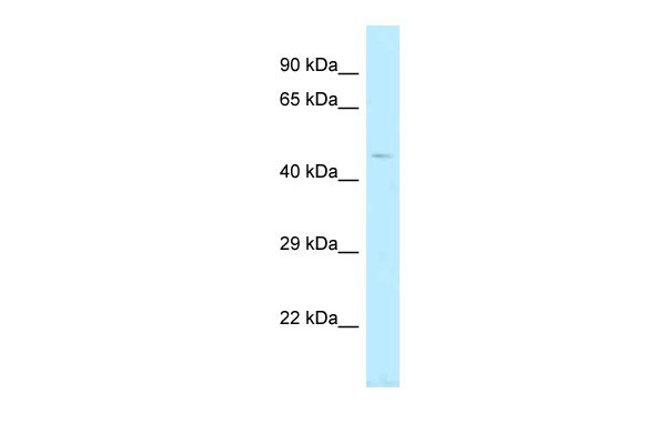
TBC1D20 antibody - C-terminal region
Rabbit Polyclonal Antibody
- SPECIFICATION
- CITATIONS
- PROTOCOLS
- BACKGROUND

Application
| WB |
|---|---|
| Primary Accession | Q96BZ9 |
| Other Accession | NM_144628, NP_653229 |
| Reactivity | Human, Mouse, Rat, Rabbit, Pig, Horse, Bovine, Guinea Pig, Dog |
| Predicted | Human, Mouse, Rat, Rabbit, Pig, Horse, Guinea Pig, Dog |
| Host | Rabbit |
| Clonality | Polyclonal |
| Calculated MW | 44kDa |
| Gene ID | 128637 |
|---|---|
| Alias Symbol | C20orf140, FLJ45119 |
| Other Names | TBC1 domain family member 20, TBC1D20, C20orf140 |
| Format | Liquid. Purified antibody supplied in 1x PBS buffer with 0.09% (w/v) sodium azide and 2% sucrose. |
| Reconstitution & Storage | Add 50 ul of distilled water. Final anti-TBC1D20 antibody concentration is 1 mg/ml in PBS buffer with 2% sucrose. For longer periods of storage, store at 20°C. Avoid repeat freeze-thaw cycles. |
| Precautions | TBC1D20 antibody - C-terminal region is for research use only and not for use in diagnostic or therapeutic procedures. |
| Name | TBC1D20 (HGNC:16133) |
|---|---|
| Synonyms | C20orf140 |
| Function | GTPase-activating protein (GAP) specific for Rab1 and Rab2 small GTPase families for which it can accelerate the intrinsic GTP hydrolysis rate by more than five orders of magnitude (PubMed:23236136). Also shows GAP activity for RAB18 GTPase (PubMed:26063829). Promotes RAB18 dissociation from the endoplasmic reticulum (ER) membrane into the cytosol, probably through stimulating RAB18 GTP-hydrolysis (PubMed:26063829). Involved in maintaining endoplasmic reticulum structure (PubMed:24891604). |
| Cellular Location | Membrane; Multi-pass membrane protein |

Thousands of laboratories across the world have published research that depended on the performance of antibodies from Abcepta to advance their research. Check out links to articles that cite our products in major peer-reviewed journals, organized by research category.
info@abcepta.com, and receive a free "I Love Antibodies" mug.

Provided below are standard protocols that you may find useful for product applications.
References
Ishibashi K.,et al.Genes Cells 14:41-52(2009).
Ota T.,et al.Nat. Genet. 36:40-45(2004).
Deloukas P.,et al.Nature 414:865-871(2001).
Mural R.J.,et al.Submitted (SEP-2005) to the EMBL/GenBank/DDBJ databases.
Sklan E.H.,et al.J. Virol. 81:11096-11105(2007).
If you have used an Abcepta product and would like to share how it has performed, please click on the "Submit Review" button and provide the requested information. Our staff will examine and post your review and contact you if needed.
If you have any additional inquiries please email technical services at tech@abcepta.com.
Ordering Information
Shipping Information
