


 Foundational characteristics of cancer include proliferation, angiogenesis, migration, evasion of apoptosis, and cellular immortality. Find key markers for these cellular processes and antibodies to detect them.
 The SUMOplot™ Analysis Program predicts and scores sumoylation sites in your protein. SUMOylation is a post-translational modification involved in various cellular processes, such as nuclear-cytosolic transport, transcriptional regulation, apoptosis, protein stability, response to stress, and progression through the cell cycle.
 The Autophagy Receptor Motif Plotter predicts and scores autophagy receptor binding sites in your protein. Identifying proteins connected to this pathway is critical to understanding the role of autophagy in physiological as well as pathological processes such as development, differentiation, neurodegenerative diseases, stress, infection, and cancer.
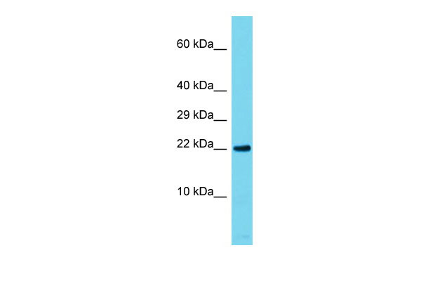
Rap1a Antibody - C-terminal region
Rabbit Polyclonal Antibody
- SPECIFICATION
- CITATIONS
- PROTOCOLS
- BACKGROUND

Application
| WB |
|---|---|
| Primary Accession | P62835 |
| Other Accession | NM_145541, NP_663516 |
| Reactivity | Human, Mouse, Rat, Rabbit, Pig, Goat, Horse, Bovine, Guinea Pig, Dog |
| Predicted | Human, Mouse, Rat, Rabbit, Pig, Goat, Horse, Bovine, Guinea Pig, Dog |
| Host | Rabbit |
| Clonality | Polyclonal |
| Calculated MW | 20kDa |
| Gene ID | 109905 |
|---|---|
| Alias Symbol | AI848598, G-22K, Krev-1, Rap1 |
| Other Names | Ras-related protein Rap-1A, Ras-related protein Krev-1, Rap1a, Krev-1 |
| Format | Liquid. Purified antibody supplied in 1x PBS buffer with 0.09% (w/v) sodium azide and 2% sucrose. |
| Reconstitution & Storage | Add 50 ul of distilled water. Final anti-Rap1a antibody concentration is 1 mg/ml in PBS buffer with 2% sucrose. For longer periods of storage, store at 20°C. Avoid repeat freeze-thaw cycles. |
| Precautions | Rap1a Antibody - C-terminal region is for research use only and not for use in diagnostic or therapeutic procedures. |
| Name | Rap1a |
|---|---|
| Synonyms | Krev-1 |
| Function | Counteracts the mitogenic function of Ras, at least partly because it can interact with Ras GAPs and RAF in a competitive manner. Together with ITGB1BP1, regulates KRIT1 localization to microtubules and membranes (By similarity). Plays a role in nerve growth factor (NGF)-induced neurite outgrowth. Plays a role in the regulation of embryonic blood vessel formation. Involved in the establishment of basal endothelial barrier function. Facilitates the progressive accumulation of CDH1 at mature desmosome junctions via cAMP-dependent signaling and its interaction with PKP3 (By similarity). May be involved in the regulation of the vascular endothelial growth factor receptor KDR expression at endothelial cell-cell junctions. |
| Cellular Location | Cell membrane; Lipid-anchor. Cell junction. Cytoplasm. Cytoplasm, perinuclear region. Early endosome. Late endosome. Note=Recruited from early endosome to late endosome compartment after nerve growth factor (NGF) stimulation. Colocalized with RAPGEF2 in the perinuclear region (By similarity). Localized with RAPGEF2 at cell-cell junctions |
| Tissue Location | Expressed in the testis and sperm midpiece (at protein level). |
Research Areas
Citations (0)

Thousands of laboratories across the world have published research that depended on the performance of antibodies from Abcepta to advance their research. Check out links to articles that cite our products in major peer-reviewed journals, organized by research category.
Submit your citation using an Abcepta antibody to
info@abcepta.com, and receive a free "I Love Antibodies" mug.
info@abcepta.com, and receive a free "I Love Antibodies" mug.

Application Protocols
Provided below are standard protocols that you may find useful for product applications.
References
Kanemura H.,et al.Biochem. Biophys. Res. Commun. 387:754-759(2009).
Abcepta welcomes feedback from its customers.
If you have used an Abcepta product and would like to share how it has performed, please click on the "Submit Review" button and provide the requested information. Our staff will examine and post your review and contact you if needed.
If you have any additional inquiries please email technical services at tech@abcepta.com.
$ 389.00
Cat# AI14958
Ordering Information
United States
AlbaniaAustraliaAustriaBelgiumBosnia & HerzegovinaBrazilBulgariaCanadaCentral AmericaChinaCroatiaCyprusCzech RepublicDenmarkEstoniaFinlandFranceGermanyGreeceHong KongHungaryIcelandIndiaIndonesiaIrelandIsraelItalyJapanLatviaLithuaniaLuxembourgMacedoniaMalaysiaMaltaMexicoNetherlandsNew ZealandNorwayPakistanPolandPortugalRomaniaSerbiaSingaporeSlovakiaSloveniaSouth AfricaSouth KoreaSpainSwedenSwitzerlandTaiwanTurkeyUnited KingdomUnited StatesVietnamWorldwideOthers
USA Headquarters
(888) 735-7227 / (858) 622-0099 or (858) 875-1900
Shipping Information
Domestic orders (in stock items)
Shipped out the same day. Orders placed after 1 PM (PST) will ship out the next business day.
International orders
Contact your local distributors
