


 Foundational characteristics of cancer include proliferation, angiogenesis, migration, evasion of apoptosis, and cellular immortality. Find key markers for these cellular processes and antibodies to detect them.
 The SUMOplot™ Analysis Program predicts and scores sumoylation sites in your protein. SUMOylation is a post-translational modification involved in various cellular processes, such as nuclear-cytosolic transport, transcriptional regulation, apoptosis, protein stability, response to stress, and progression through the cell cycle.
 The Autophagy Receptor Motif Plotter predicts and scores autophagy receptor binding sites in your protein. Identifying proteins connected to this pathway is critical to understanding the role of autophagy in physiological as well as pathological processes such as development, differentiation, neurodegenerative diseases, stress, infection, and cancer.
RCN3 Antibody - C-terminal region
Rabbit Polyclonal Antibody
- SPECIFICATION
- CITATIONS
- PROTOCOLS
- BACKGROUND

Application
| WB |
|---|---|
| Primary Accession | Q96D15 |
| Other Accession | NM_020650, NP_065701 |
| Reactivity | Human, Mouse, Rat, Rabbit, Pig, Horse, Bovine, Guinea Pig, Dog |
| Predicted | Human, Mouse, Rat, Rabbit, Pig, Horse, Bovine, Guinea Pig, Dog |
| Host | Rabbit |
| Clonality | Polyclonal |
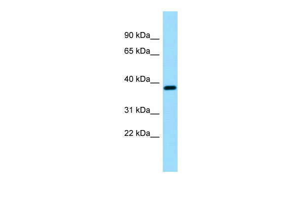
| Calculated MW | 35kDa |
| Gene ID | 57333 |
|---|---|
| Alias Symbol | RLP49 |
| Other Names | Reticulocalbin-3, EF-hand calcium-binding protein RLP49, RCN3 |
| Format | Liquid. Purified antibody supplied in 1x PBS buffer with 0.09% (w/v) sodium azide and 2% sucrose. |
| Reconstitution & Storage | Add 50 ul of distilled water. Final anti-RCN3 antibody concentration is 1 mg/ml in PBS buffer with 2% sucrose. For longer periods of storage, store at 20°C. Avoid repeat freeze-thaw cycles. |
| Precautions | RCN3 Antibody - C-terminal region is for research use only and not for use in diagnostic or therapeutic procedures. |
| Name | RCN3 (HGNC:21145) |
|---|---|
| Function | Probable molecular chaperone assisting protein biosynthesis and transport in the endoplasmic reticulum (PubMed:16433634, PubMed:28939891). Required for the proper biosynthesis and transport of pulmonary surfactant-associated protein A/SP-A, pulmonary surfactant- associated protein D/SP-D and the lipid transporter ABCA3 (By similarity). By regulating both the proper expression and the degradation through the endoplasmic reticulum-associated protein degradation pathway of these proteins plays a crucial role in pulmonary surfactant homeostasis (By similarity). Has an anti-fibrotic activity by negatively regulating the secretion of type I and type III collagens (PubMed:28939891). This calcium-binding protein also transiently associates with immature PCSK6 and regulates its secretion (PubMed:16433634). |
| Cellular Location | Endoplasmic reticulum lumen |
| Tissue Location | Widely expressed.. |

Thousands of laboratories across the world have published research that depended on the performance of antibodies from Abcepta to advance their research. Check out links to articles that cite our products in major peer-reviewed journals, organized by research category.
info@abcepta.com, and receive a free "I Love Antibodies" mug.

Provided below are standard protocols that you may find useful for product applications.
References
Liu F.,et al.Submitted (DEC-2002) to the EMBL/GenBank/DDBJ databases.
Hu R.-M.,et al.Proc. Natl. Acad. Sci. U.S.A. 97:9543-9548(2000).
Clark H.F.,et al.Genome Res. 13:2265-2270(2003).
Chen R.,et al.J. Proteome Res. 8:651-661(2009).
Burkard T.R.,et al.BMC Syst. Biol. 5:17-17(2011).
If you have used an Abcepta product and would like to share how it has performed, please click on the "Submit Review" button and provide the requested information. Our staff will examine and post your review and contact you if needed.
If you have any additional inquiries please email technical services at tech@abcepta.com.
Ordering Information
Shipping Information
