


 Foundational characteristics of cancer include proliferation, angiogenesis, migration, evasion of apoptosis, and cellular immortality. Find key markers for these cellular processes and antibodies to detect them.
 The SUMOplot™ Analysis Program predicts and scores sumoylation sites in your protein. SUMOylation is a post-translational modification involved in various cellular processes, such as nuclear-cytosolic transport, transcriptional regulation, apoptosis, protein stability, response to stress, and progression through the cell cycle.
 The Autophagy Receptor Motif Plotter predicts and scores autophagy receptor binding sites in your protein. Identifying proteins connected to this pathway is critical to understanding the role of autophagy in physiological as well as pathological processes such as development, differentiation, neurodegenerative diseases, stress, infection, and cancer.
RCN3 Antibody (Center)
Affinity Purified Rabbit Polyclonal Antibody (Pab)
- SPECIFICATION
- CITATIONS: 1
- PROTOCOLS
- BACKGROUND

Application
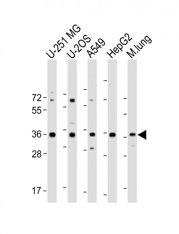
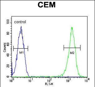
| WB, FC, E |
|---|---|
| Primary Accession | Q96D15 |
| Other Accession | Q8BH97, Q2KJ39, NP_065701.2, I6L9G5 |
| Reactivity | Human, Mouse |
| Predicted | Bovine, Rat |
| Host | Rabbit |
| Clonality | Polyclonal |
| Isotype | Rabbit IgG |
| Calculated MW | 37493 Da |
| Antigen Region | 134-163 aa |
| Gene ID | 57333 |
|---|---|
| Other Names | Reticulocalbin-3, EF-hand calcium-binding protein RLP49, RCN3 |
| Target/Specificity | This RCN3 antibody is generated from rabbits immunized with a KLH conjugated synthetic peptide between 134-163 amino acids from the Central region of human RCN3. |
| Dilution | WB~~1:2000 FC~~1:10~50 E~~Use at an assay dependent concentration. |
| Format | Purified polyclonal antibody supplied in PBS with 0.09% (W/V) sodium azide. This antibody is purified through a protein A column, followed by peptide affinity purification. |
| Storage | Maintain refrigerated at 2-8°C for up to 2 weeks. For long term storage store at -20°C in small aliquots to prevent freeze-thaw cycles. |
| Precautions | RCN3 Antibody (Center) is for research use only and not for use in diagnostic or therapeutic procedures. |
| Name | RCN3 (HGNC:21145) |
|---|---|
| Function | Probable molecular chaperone assisting protein biosynthesis and transport in the endoplasmic reticulum (PubMed:16433634, PubMed:28939891). Required for the proper biosynthesis and transport of pulmonary surfactant-associated protein A/SP-A, pulmonary surfactant- associated protein D/SP-D and the lipid transporter ABCA3 (By similarity). By regulating both the proper expression and the degradation through the endoplasmic reticulum-associated protein degradation pathway of these proteins plays a crucial role in pulmonary surfactant homeostasis (By similarity). Has an anti-fibrotic activity by negatively regulating the secretion of type I and type III collagens (PubMed:28939891). This calcium-binding protein also transiently associates with immature PCSK6 and regulates its secretion (PubMed:16433634). |
| Cellular Location | Endoplasmic reticulum lumen |
| Tissue Location | Widely expressed.. |

Provided below are standard protocols that you may find useful for product applications.
Background
Calcipressin 3 inhibits calcineurin dependent transcriptional responses by binding to the catalytic domain of calcineurin A, and may play a role during central nervous system development. Highest expression occurs in heart, skeletal muscle kidney, liver and peripheral blood leukocytes.
References
Tsuji, A., et al. Biochem. J. 396(1):51-59(2006)
Clark, H.F., et al. Genome Res. 13(10):2265-2270(2003)
Hu, R.M., et al. Proc. Natl. Acad. Sci. U.S.A. 97(17):9543-9548(2000)
If you have used an Abcepta product and would like to share how it has performed, please click on the "Submit Review" button and provide the requested information. Our staff will examine and post your review and contact you if needed.
If you have any additional inquiries please email technical services at tech@abcepta.com.
Ordering Information
Shipping Information
