


 Foundational characteristics of cancer include proliferation, angiogenesis, migration, evasion of apoptosis, and cellular immortality. Find key markers for these cellular processes and antibodies to detect them.
 The SUMOplot™ Analysis Program predicts and scores sumoylation sites in your protein. SUMOylation is a post-translational modification involved in various cellular processes, such as nuclear-cytosolic transport, transcriptional regulation, apoptosis, protein stability, response to stress, and progression through the cell cycle.
 The Autophagy Receptor Motif Plotter predicts and scores autophagy receptor binding sites in your protein. Identifying proteins connected to this pathway is critical to understanding the role of autophagy in physiological as well as pathological processes such as development, differentiation, neurodegenerative diseases, stress, infection, and cancer.
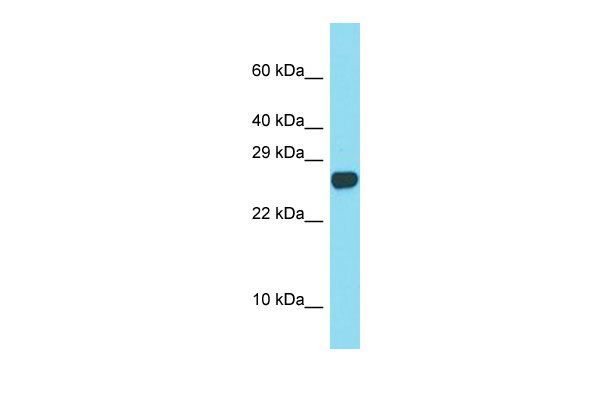
Spaca3 Antibody - C-terminal region
Rabbit Polyclonal Antibody
- SPECIFICATION
- CITATIONS
- PROTOCOLS
- BACKGROUND

Application
| WB |
|---|---|
| Primary Accession | Q9D9X8 |
| Reactivity | Human, Mouse, Rat, Rabbit, Pig, Goat, Sheep, Horse, Bovine, Dog |
| Predicted | Human, Mouse, Rat, Rabbit, Pig, Goat, Sheep, Horse, Bovine, Dog |
| Host | Rabbit |
| Clonality | Polyclonal |
| Calculated MW | 24kDa |
| Gene ID | 75622 |
|---|---|
| Other Names | Sperm acrosome membrane-associated protein 3, Lysozyme-like protein 3, Sperm lysozyme-like protein 1, mSLLP1, Sperm acrosome membrane-associated protein 3, membrane form, Sperm acrosome membrane-associated protein 3, processed form, Spaca3, Lyc3, Lyzl3, Sllp1 |
| Format | Liquid. Purified antibody supplied in 1x PBS buffer with 0.09% (w/v) sodium azide and 2% sucrose. |
| Reconstitution & Storage | Add 50 ul of distilled water. Final anti-Spaca3 antibody concentration is 1 mg/ml in PBS buffer with 2% sucrose. For longer periods of storage, store at 20°C. Avoid repeat freeze-thaw cycles. |
| Precautions | Spaca3 Antibody - C-terminal region is for research use only and not for use in diagnostic or therapeutic procedures. |
| Name | Spaca3 |
|---|---|
| Synonyms | Lyc3, Lyzl3, Sllp1 |
| Function | Sperm surface membrane protein that may be involved in sperm- egg plasma membrane adhesion and fusion during fertilization. It could be a potential receptor for the egg oligosaccharide residue N- acetylglucosamine, which is present in the extracellular matrix over the egg plasma membrane. The processed form has no detectable bacteriolytic activity in vitro (By similarity). |
| Cellular Location | Cytoplasmic vesicle, secretory vesicle, acrosome membrane; Single-pass type II membrane protein. Note=Anterior acrosome in non- capacitated spermatozoa and retained in the equatorial segment and in the luminal face of both the inner and outer acrosomal membranes following capacitation and the acrosome reaction |
| Tissue Location | The processed form is expressed in sperm (at protein level). Expressed strongly in testis and epididymis and weakly in pancreas. |

Thousands of laboratories across the world have published research that depended on the performance of antibodies from Abcepta to advance their research. Check out links to articles that cite our products in major peer-reviewed journals, organized by research category.
info@abcepta.com, and receive a free "I Love Antibodies" mug.

Provided below are standard protocols that you may find useful for product applications.
References
Zhang K.,et al.Biol. Reprod. 73:1064-1071(2005).
Carninci P.,et al.Science 309:1559-1563(2005).
Church D.M.,et al.PLoS Biol. 7:E1000112-E1000112(2009).
Herrero M.B.,et al.Dev. Biol. 284:126-142(2005).
Sachdev M.,et al.Dev. Biol. 363:40-51(2012).
If you have used an Abcepta product and would like to share how it has performed, please click on the "Submit Review" button and provide the requested information. Our staff will examine and post your review and contact you if needed.
If you have any additional inquiries please email technical services at tech@abcepta.com.
Ordering Information
Other Products
Shipping Information
