


 Foundational characteristics of cancer include proliferation, angiogenesis, migration, evasion of apoptosis, and cellular immortality. Find key markers for these cellular processes and antibodies to detect them.
 The SUMOplot™ Analysis Program predicts and scores sumoylation sites in your protein. SUMOylation is a post-translational modification involved in various cellular processes, such as nuclear-cytosolic transport, transcriptional regulation, apoptosis, protein stability, response to stress, and progression through the cell cycle.
 The Autophagy Receptor Motif Plotter predicts and scores autophagy receptor binding sites in your protein. Identifying proteins connected to this pathway is critical to understanding the role of autophagy in physiological as well as pathological processes such as development, differentiation, neurodegenerative diseases, stress, infection, and cancer.
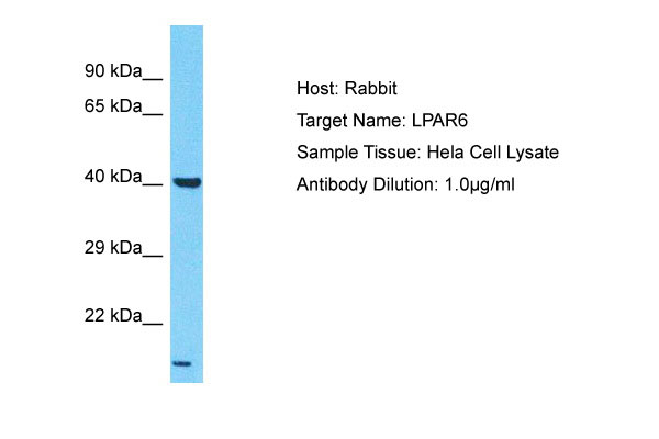
LPAR6 Antibody - N-terminal region
Rabbit Polyclonal Antibody
- SPECIFICATION
- CITATIONS
- PROTOCOLS
- BACKGROUND

Application
| WB |
|---|---|
| Primary Accession | P43657 |
| Other Accession | NM_005767, NP_005758 |
| Reactivity | Human, Mouse, Rat, Rabbit, Pig, Sheep, Horse, Bovine, Dog |
| Predicted | Human, Mouse, Rat, Rabbit, Pig, Sheep, Horse, Bovine, Dog |
| Host | Rabbit |
| Clonality | Polyclonal |
| Calculated MW | 37kDa |
| Gene ID | 10161 |
|---|---|
| Alias Symbol | ARWH1, LAH3, P2RY5, P2Y5 |
| Other Names | Lysophosphatidic acid receptor 6, LPA receptor 6, LPA-6, Oleoyl-L-alpha-lysophosphatidic acid receptor, P2Y purinoceptor 5, P2Y5, Purinergic receptor 5, RB intron encoded G-protein coupled receptor, LPAR6, P2RY5 |
| Format | Liquid. Purified antibody supplied in 1x PBS buffer with 0.09% (w/v) sodium azide and 2% sucrose. |
| Reconstitution & Storage | Add 50 &mu, l of distilled water. Final Anti-LPAR6 antibody concentration is 1 mg/ml in PBS buffer with 2% sucrose. For longer periods of storage, store at -20°C. Avoid repeat freeze-thaw cycles. |
| Precautions | LPAR6 Antibody - N-terminal region is for research use only and not for use in diagnostic or therapeutic procedures. |
| Name | LPAR6 |
|---|---|
| Synonyms | P2RY5 |
| Function | Binds to oleoyl-L-alpha-lysophosphatidic acid (LPA). Intracellular cAMP is involved in the receptor activation. Important for the maintenance of hair growth and texture. |
| Cellular Location | Cell membrane; Multi-pass membrane protein |
| Tissue Location | Expressed ubiquitously, including in skin and hair follicle cells. Detected in both Henle's and Huxley's layers of the inner root sheath of the hair follicle and in suprabasal layers of the epidermis (at protein level). Expressed at low levels in peripheral blood leukocytes. |

Thousands of laboratories across the world have published research that depended on the performance of antibodies from Abcepta to advance their research. Check out links to articles that cite our products in major peer-reviewed journals, organized by research category.
info@abcepta.com, and receive a free "I Love Antibodies" mug.

Provided below are standard protocols that you may find useful for product applications.
References
Toguchida J.,et al.Genomics 17:535-543(1993).
Herzog H.,et al.Genome Res. 6:858-861(1996).
Bohm S.K.,et al.Submitted (JUL-1997) to the EMBL/GenBank/DDBJ databases.
Bechtel S.,et al.BMC Genomics 8:399-399(2007).
Ota T.,et al.Nat. Genet. 36:40-45(2004).
If you have used an Abcepta product and would like to share how it has performed, please click on the "Submit Review" button and provide the requested information. Our staff will examine and post your review and contact you if needed.
If you have any additional inquiries please email technical services at tech@abcepta.com.
Ordering Information
Other Products
Shipping Information
