


 Foundational characteristics of cancer include proliferation, angiogenesis, migration, evasion of apoptosis, and cellular immortality. Find key markers for these cellular processes and antibodies to detect them.
 The SUMOplot™ Analysis Program predicts and scores sumoylation sites in your protein. SUMOylation is a post-translational modification involved in various cellular processes, such as nuclear-cytosolic transport, transcriptional regulation, apoptosis, protein stability, response to stress, and progression through the cell cycle.
 The Autophagy Receptor Motif Plotter predicts and scores autophagy receptor binding sites in your protein. Identifying proteins connected to this pathway is critical to understanding the role of autophagy in physiological as well as pathological processes such as development, differentiation, neurodegenerative diseases, stress, infection, and cancer.
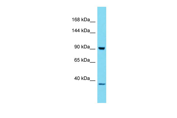
HKDC1 Antibody - C-terminal region
Rabbit Polyclonal Antibody
- SPECIFICATION
- CITATIONS
- PROTOCOLS
- BACKGROUND

Application
| WB |
|---|---|
| Primary Accession | Q2TB90 |
| Other Accession | NM_025130, NP_079406 |
| Reactivity | Human, Mouse, Rat, Rabbit, Pig, Horse, Bovine, Dog |
| Predicted | Human, Mouse, Rat, Rabbit, Pig, Horse, Bovine, Dog |
| Host | Rabbit |
| Clonality | Polyclonal |
| Calculated MW | 102kDa |
| Gene ID | 80201 |
|---|---|
| Other Names | Putative hexokinase HKDC1, 2.7.1.1, Hexokinase domain-containing protein 1, HKDC1 |
| Format | Liquid. Purified antibody supplied in 1x PBS buffer with 0.09% (w/v) sodium azide and 2% sucrose. |
| Reconstitution & Storage | Add 50 ul of distilled water. Final anti-HKDC1 antibody concentration is 1 mg/ml in PBS buffer with 2% sucrose. For longer periods of storage, store at 20°C. Avoid repeat freeze-thaw cycles. |
| Precautions | HKDC1 Antibody - C-terminal region is for research use only and not for use in diagnostic or therapeutic procedures. |
| Name | HKDC1 (HGNC:23302) |
|---|---|
| Function | Catalyzes the phosphorylation of hexose to hexose 6- phosphate, although at very low level compared to other hexokinases (PubMed:30517626). Has low glucose phosphorylating activity compared to other hexokinases (PubMed:30517626). Involved in glucose homeostasis and hepatic lipid accumulation. Required to maintain whole-body glucose homeostasis during pregnancy; however additional evidences are required to confirm this role (By similarity). |
| Cellular Location | Cytoplasm. Mitochondrion membrane; Peripheral membrane protein. Photoreceptor inner segment {ECO:0000250|UniProtKB:Q91W97}. Note=The mitochondrial-binding peptide (MBP) region promotes association with the mitochondrion |
| Tissue Location | Widely expressed (PubMed:27459389, PubMed:29401404). Highly expressed in the brush border, surface epithelium and the myenteric plexus of the small and large intestines; the acinar centrocytes and interlobular ducts of the pancreas; and the alveolar macrophages in the lungs (at protein level) (PubMed:29401404) Present at moderate level in the thyroid follicular epithelium (at protein level) (PubMed:29401404). |

Thousands of laboratories across the world have published research that depended on the performance of antibodies from Abcepta to advance their research. Check out links to articles that cite our products in major peer-reviewed journals, organized by research category.
info@abcepta.com, and receive a free "I Love Antibodies" mug.

Provided below are standard protocols that you may find useful for product applications.
References
Deloukas P.,et al.Nature 429:375-381(2004).
Ota T.,et al.Nat. Genet. 36:40-45(2004).
Bechtel S.,et al.BMC Genomics 8:399-399(2007).
Burkard T.R.,et al.BMC Syst. Biol. 5:17-17(2011).
If you have used an Abcepta product and would like to share how it has performed, please click on the "Submit Review" button and provide the requested information. Our staff will examine and post your review and contact you if needed.
If you have any additional inquiries please email technical services at tech@abcepta.com.
Ordering Information
Other Products
Shipping Information
