


 Foundational characteristics of cancer include proliferation, angiogenesis, migration, evasion of apoptosis, and cellular immortality. Find key markers for these cellular processes and antibodies to detect them.
 The SUMOplot™ Analysis Program predicts and scores sumoylation sites in your protein. SUMOylation is a post-translational modification involved in various cellular processes, such as nuclear-cytosolic transport, transcriptional regulation, apoptosis, protein stability, response to stress, and progression through the cell cycle.
 The Autophagy Receptor Motif Plotter predicts and scores autophagy receptor binding sites in your protein. Identifying proteins connected to this pathway is critical to understanding the role of autophagy in physiological as well as pathological processes such as development, differentiation, neurodegenerative diseases, stress, infection, and cancer.
CCDC66 Antibody - N-terminal region
Rabbit Polyclonal Antibody
- SPECIFICATION
- CITATIONS
- PROTOCOLS
- BACKGROUND

Application
| WB |
|---|---|
| Primary Accession | A2RUB6 |
| Other Accession | NM_001012506, NP_001012524 |
| Reactivity | Human, Horse, Bovine, Dog |
| Predicted | Human, Horse, Bovine, Dog |
| Host | Rabbit |
| Clonality | Polyclonal |
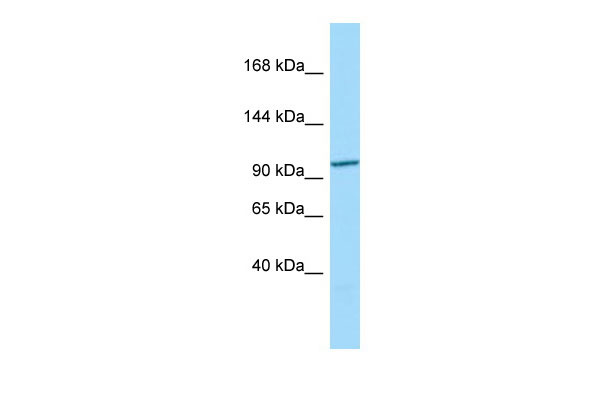
| Calculated MW | 91kDa |
| Gene ID | 285331 |
|---|---|
| Other Names | Coiled-coil domain-containing protein 66, CCDC66 |
| Format | Liquid. Purified antibody supplied in 1x PBS buffer with 0.09% (w/v) sodium azide and 2% sucrose. |
| Reconstitution & Storage | Add 50 ul of distilled water. Final anti-CCDC66 antibody concentration is 1 mg/ml in PBS buffer with 2% sucrose. For longer periods of storage, store at 20°C. Avoid repeat freeze-thaw cycles. |
| Precautions | CCDC66 Antibody - N-terminal region is for research use only and not for use in diagnostic or therapeutic procedures. |
| Name | CCDC66 (HGNC:27709) |
|---|---|
| Function | Microtubule-binding protein required for ciliogenesis (PubMed:28235840). May function in ciliogenesis by mediating the transport of proteins like BBS4 to the cilium, but also through the organization of the centriolar satellites (PubMed:28235840). Required for the assembly of signaling-competent cilia with proper structure and length (PubMed:36606424). Mediates this function in part by regulating transition zone assembly and basal body recruitment of the IFT-B complex (PubMed:36606424). Cooperates with the ciliopathy proteins CSPP1 and CEP104 during cilium length regulation (PubMed:36606424). Plays two important roles during cell division (PubMed:35849559). First, is required for mitotic progression via regulation of spindle assembly, organization and orientation, levels of spindle microtubules (MTs), kinetochore-fiber integrity, and chromosome alignment (PubMed:35849559). Second, functions during cytokinesis in part by regulating assembly and organization of central spindle and midbody MTs (PubMed:35849559). Plays a role in retina morphogenesis and/or homeostasis (By similarity). |
| Cellular Location | Cytoplasm, cytoskeleton, microtubule organizing center, centrosome. Cytoplasm, cytoskeleton, microtubule organizing center, centrosome, centriolar satellite. Cell projection, cilium. Cytoplasm, cytoskeleton, cilium basal body. Cytoplasm, cytoskeleton, cilium axoneme. Photoreceptor inner segment. Cell projection, cilium, photoreceptor outer segment. Cytoplasm, cytoskeleton, spindle. Midbody. Note=Restricted to the centrosomes and the spindle microtubules during mitosis (PubMed:28235840). Enriched in the inner segment of the photoreceptor (PubMed:19777273) |
| Tissue Location | Widely expressed (at protein level) (PubMed:28235840). Expressed in retina, mainly in photoreceptors but also in outer plexiform and ganglion cell layers (at protein level) (PubMed:19777273). |
Citations (0)

Thousands of laboratories across the world have published research that depended on the performance of antibodies from Abcepta to advance their research. Check out links to articles that cite our products in major peer-reviewed journals, organized by research category.
Submit your citation using an Abcepta antibody to
info@abcepta.com, and receive a free "I Love Antibodies" mug.
info@abcepta.com, and receive a free "I Love Antibodies" mug.

Application Protocols
Provided below are standard protocols that you may find useful for product applications.
Abcepta welcomes feedback from its customers.
If you have used an Abcepta product and would like to share how it has performed, please click on the "Submit Review" button and provide the requested information. Our staff will examine and post your review and contact you if needed.
If you have any additional inquiries please email technical services at tech@abcepta.com.
$ 389.00
Cat# AI15582
Ordering Information
United States
AlbaniaAustraliaAustriaBelgiumBosnia & HerzegovinaBrazilBulgariaCanadaCentral AmericaChinaCroatiaCyprusCzech RepublicDenmarkEstoniaFinlandFranceGermanyGreeceHong KongHungaryIcelandIndiaIndonesiaIrelandIsraelItalyJapanLatviaLithuaniaLuxembourgMacedoniaMalaysiaMaltaMexicoNetherlandsNew ZealandNorwayPakistanPolandPortugalRomaniaSerbiaSingaporeSlovakiaSloveniaSouth AfricaSouth KoreaSpainSwedenSwitzerlandTaiwanTurkeyUnited KingdomUnited StatesVietnamWorldwideOthers
USA Headquarters
(888) 735-7227 / (858) 622-0099 or (858) 875-1900
Shipping Information
Domestic orders (in stock items)
Shipped out the same day. Orders placed after 1 PM (PST) will ship out the next business day.
International orders
Contact your local distributors
