


 Foundational characteristics of cancer include proliferation, angiogenesis, migration, evasion of apoptosis, and cellular immortality. Find key markers for these cellular processes and antibodies to detect them.
 The SUMOplot™ Analysis Program predicts and scores sumoylation sites in your protein. SUMOylation is a post-translational modification involved in various cellular processes, such as nuclear-cytosolic transport, transcriptional regulation, apoptosis, protein stability, response to stress, and progression through the cell cycle.
 The Autophagy Receptor Motif Plotter predicts and scores autophagy receptor binding sites in your protein. Identifying proteins connected to this pathway is critical to understanding the role of autophagy in physiological as well as pathological processes such as development, differentiation, neurodegenerative diseases, stress, infection, and cancer.
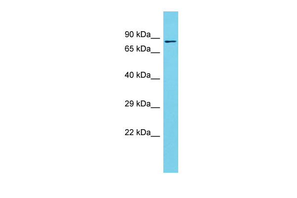
SPATA5L1 Antibody - C-terminal region
Rabbit Polyclonal Antibody
- SPECIFICATION
- CITATIONS
- PROTOCOLS
- BACKGROUND

Application
| WB |
|---|---|
| Primary Accession | Q9BVQ7 |
| Other Accession | NM_024063, NP_076968 |
| Reactivity | Human, Rat, Rabbit, Pig, Bovine, Dog |
| Predicted | Human, Rat, Rabbit, Pig, Bovine, Dog |
| Host | Rabbit |
| Clonality | Polyclonal |
| Calculated MW | 80kDa |
| Gene ID | 79029 |
|---|---|
| Other Names | Spermatogenesis-associated protein 5-like protein 1, SPATA5L1 |
| Format | Liquid. Purified antibody supplied in 1x PBS buffer with 0.09% (w/v) sodium azide and 2% sucrose. |
| Reconstitution & Storage | Add 50 ul of distilled water. Final anti-SPATA5L1 antibody concentration is 1 mg/ml in PBS buffer with 2% sucrose. For longer periods of storage, store at 20°C. Avoid repeat freeze-thaw cycles. |
| Precautions | SPATA5L1 Antibody - C-terminal region is for research use only and not for use in diagnostic or therapeutic procedures. |
| Name | AFG2B (HGNC:28762) |
|---|---|
| Function | ATP-dependent chaperone part of the 55LCC heterohexameric ATPase complex which is chromatin-associated and promotes replisome proteostasis to maintain replication fork progression and genome stability. Required for replication fork progression, sister chromatid cohesion, and chromosome stability. The ATPase activity is specifically enhanced by replication fork DNA and is coupled to cysteine protease- dependent cleavage of replisome substrates in response to replication fork damage. Uses ATPase activity to process replisome substrates in S- phase, facilitating their proteolytic turnover from chromatin to ensure DNA replication and mitotic fidelity (PubMed:38554706). Plays an essential role in the cytoplasmic maturation steps of pre-60S ribosomal particles by promoting the release of shuttling protein RSL24D1/RLP24 from the pre-ribosomal particles (PubMed:35354024). |
| Cellular Location | Cytoplasm. Cytoplasm, cytoskeleton, spindle. Nucleus {ECO:0000250|UniProtKB:D4A2B7} |
| Tissue Location | Expressed in both neurons and glia during embryonic and adult stages of brain development. |

Thousands of laboratories across the world have published research that depended on the performance of antibodies from Abcepta to advance their research. Check out links to articles that cite our products in major peer-reviewed journals, organized by research category.
info@abcepta.com, and receive a free "I Love Antibodies" mug.

Provided below are standard protocols that you may find useful for product applications.
References
Ota T.,et al.Nat. Genet. 36:40-45(2004).
Zody M.C.,et al.Nature 440:671-675(2006).
Burkard T.R.,et al.BMC Syst. Biol. 5:17-17(2011).
Van Damme P.,et al.Proc. Natl. Acad. Sci. U.S.A. 109:12449-12454(2012).
If you have used an Abcepta product and would like to share how it has performed, please click on the "Submit Review" button and provide the requested information. Our staff will examine and post your review and contact you if needed.
If you have any additional inquiries please email technical services at tech@abcepta.com.
Ordering Information
Shipping Information
