


 Foundational characteristics of cancer include proliferation, angiogenesis, migration, evasion of apoptosis, and cellular immortality. Find key markers for these cellular processes and antibodies to detect them.
 The SUMOplot™ Analysis Program predicts and scores sumoylation sites in your protein. SUMOylation is a post-translational modification involved in various cellular processes, such as nuclear-cytosolic transport, transcriptional regulation, apoptosis, protein stability, response to stress, and progression through the cell cycle.
 The Autophagy Receptor Motif Plotter predicts and scores autophagy receptor binding sites in your protein. Identifying proteins connected to this pathway is critical to understanding the role of autophagy in physiological as well as pathological processes such as development, differentiation, neurodegenerative diseases, stress, infection, and cancer.
RGS1 Antibody - N-terminal region
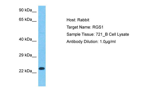
Rabbit Polyclonal Antibody
- SPECIFICATION
- CITATIONS
- PROTOCOLS
- BACKGROUND

Application
| WB |
|---|---|
| Primary Accession | Q08116 |
| Other Accession | NP_002913 |
| Reactivity | Human |
| Host | Rabbit |
| Clonality | Polyclonal |
| Calculated MW | 22kDa |
| Gene ID | 5996 |
|---|---|
| Alias Symbol | RGS1, 1R20, BL34, IER1, |
| Other Names | Regulator of G-protein signaling 1, RGS1, B-cell activation protein BL34, Early response protein 1R20, RGS1, 1R20, BL34, IER1 |
| Format | Liquid. Purified antibody supplied in 1x PBS buffer with 0.09% (w/v) sodium azide and 2% sucrose. |
| Reconstitution & Storage | Add 50 &mu, l of distilled water. Final Anti-RGS1 antibody concentration is 1 mg/ml in PBS buffer with 2% sucrose. For longer periods of storage, store at -20°C. Avoid repeat freeze-thaw cycles. |
| Precautions | RGS1 Antibody - N-terminal region is for research use only and not for use in diagnostic or therapeutic procedures. |
| Name | RGS1 |
|---|---|
| Synonyms | 1R20, BL34, IER1 |
| Function | Regulates G protein-coupled receptor signaling cascades, including signaling downstream of the N-formylpeptide chemoattractant receptors and leukotriene receptors (PubMed:10480894). Inhibits B cell chemotaxis toward CXCL12 (By similarity). Inhibits signal transduction by increasing the GTPase activity of G protein alpha subunits thereby driving them into their inactive GDP-bound form (PubMed:10480894, PubMed:18434541). |
| Cellular Location | Cell membrane; Peripheral membrane protein; Cytoplasmic side. Cytoplasm, cytosol |
| Tissue Location | Detected in peripheral blood monocytes (PubMed:10480894). Expression is relatively low in B-cells and chronic lymphocytic leukemia B-cells; however, in other types of malignant B- cell such as non-Hodgkin lymphoma and hairy cell leukemia, expression is constitutively high (PubMed:8473738). |

Thousands of laboratories across the world have published research that depended on the performance of antibodies from Abcepta to advance their research. Check out links to articles that cite our products in major peer-reviewed journals, organized by research category.
info@abcepta.com, and receive a free "I Love Antibodies" mug.

Provided below are standard protocols that you may find useful for product applications.
Background
Inhibits signal transduction by increasing the GTPase activity of G protein alpha subunits thereby driving them into their inactive GDP-bound form. This protein may be involved in the regulation of B-cell activation and proliferation.
References
Ota T.,et al.Nat. Genet. 36:40-45(2004).
Gregory S.G.,et al.Nature 441:315-321(2006).
Hong J.X.,et al.J. Immunol. 150:3895-3904(1993).
Newton J.S.,et al.Biochim. Biophys. Acta 1216:314-316(1993).
Puhl H.L. III,et al.Submitted (MAR-2002) to the EMBL/GenBank/DDBJ databases.
If you have used an Abcepta product and would like to share how it has performed, please click on the "Submit Review" button and provide the requested information. Our staff will examine and post your review and contact you if needed.
If you have any additional inquiries please email technical services at tech@abcepta.com.
Ordering Information
Other Products
Shipping Information
