


 Foundational characteristics of cancer include proliferation, angiogenesis, migration, evasion of apoptosis, and cellular immortality. Find key markers for these cellular processes and antibodies to detect them.
 The SUMOplot™ Analysis Program predicts and scores sumoylation sites in your protein. SUMOylation is a post-translational modification involved in various cellular processes, such as nuclear-cytosolic transport, transcriptional regulation, apoptosis, protein stability, response to stress, and progression through the cell cycle.
 The Autophagy Receptor Motif Plotter predicts and scores autophagy receptor binding sites in your protein. Identifying proteins connected to this pathway is critical to understanding the role of autophagy in physiological as well as pathological processes such as development, differentiation, neurodegenerative diseases, stress, infection, and cancer.
NR2E1 / TLX Antibody (N-Terminus)
Rabbit Polyclonal Antibody
- SPECIFICATION
- CITATIONS
- PROTOCOLS
- BACKGROUND

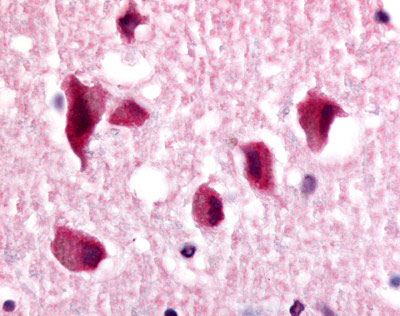
Application
| IHC-P |
|---|---|
| Primary Accession | Q9Y466 |
| Reactivity | Human, Rabbit, Monkey, Pig, Horse, Bovine, Dog |
| Host | Rabbit |
| Clonality | Polyclonal |
| Calculated MW | 43kDa |
| Dilution | IHC-P (5.5 µg/ml) |
| Gene ID | 7101 |
|---|---|
| Other Names | Nuclear receptor subfamily 2 group E member 1, Nuclear receptor TLX, Protein tailless homolog, Tll, hTll, NR2E1, TLX |
| Target/Specificity | Human NR2E1. BLAST analysis of the peptide immunogen showed no homology with other human proteins. |
| Reconstitution & Storage | Long term: -70°C; Short term: +4°C |
| Precautions | NR2E1 / TLX Antibody (N-Terminus) is for research use only and not for use in diagnostic or therapeutic procedures. |
| Name | NR2E1 |
|---|---|
| Synonyms | TLX |
| Function | Orphan receptor that binds DNA as a monomer to hormone response elements (HRE) containing an extended core motif half-site sequence 5'-AAGGTCA-3' in which the 5' flanking nucleotides participate in determining receptor specificity (By similarity). May be required to pattern anterior brain differentiation. Involved in the regulation of retinal development and essential for vision. During retinogenesis, regulates PTEN-Cyclin D expression via binding to the promoter region of PTEN and suppressing its activity (By similarity). May be involved in retinoic acid receptor (RAR) regulation in retinal cells. |
| Cellular Location | Nucleus {ECO:0000255|PROSITE-ProRule:PRU00407}. |
| Tissue Location | Brain specific. Present in all brain sections tested, highest levels in the caudate nucleus and hippocampus, weakest levels in the thalamus. |

Thousands of laboratories across the world have published research that depended on the performance of antibodies from Abcepta to advance their research. Check out links to articles that cite our products in major peer-reviewed journals, organized by research category.
info@abcepta.com, and receive a free "I Love Antibodies" mug.

Provided below are standard protocols that you may find useful for product applications.
Background
Orphan receptor that binds DNA as a monomer to hormone response elements (HRE) containing an extended core motif half- site sequence 5'-AAGGTCA-3' in which the 5' flanking nucleotides participate in determining receptor specificity (By similarity). May be required to pattern anterior brain differentiation. Involved in the regulation of retinal development and essential for vision. During retinogenesis, regulates PTEN-Cyclin D expression via binding to the promoter region of PTEN and suppressing its activity (By similarity). May be involved in retinoic acic receptor (RAR) regulation in retinal cells.
References
Jackson A.,et al.Genomics 50:34-43(1998).
Kobayashi M.,et al.Mol. Cell. Biol. 20:8731-8739(2000).
Hua F.,et al.Submitted (AUG-2001) to the EMBL/GenBank/DDBJ databases.
Ota T.,et al.Nat. Genet. 36:40-45(2004).
Mungall A.J.,et al.Nature 425:805-811(2003).
If you have used an Abcepta product and would like to share how it has performed, please click on the "Submit Review" button and provide the requested information. Our staff will examine and post your review and contact you if needed.
If you have any additional inquiries please email technical services at tech@abcepta.com.
Ordering Information
Other Products
Shipping Information
