


 Foundational characteristics of cancer include proliferation, angiogenesis, migration, evasion of apoptosis, and cellular immortality. Find key markers for these cellular processes and antibodies to detect them.
 The SUMOplot™ Analysis Program predicts and scores sumoylation sites in your protein. SUMOylation is a post-translational modification involved in various cellular processes, such as nuclear-cytosolic transport, transcriptional regulation, apoptosis, protein stability, response to stress, and progression through the cell cycle.
 The Autophagy Receptor Motif Plotter predicts and scores autophagy receptor binding sites in your protein. Identifying proteins connected to this pathway is critical to understanding the role of autophagy in physiological as well as pathological processes such as development, differentiation, neurodegenerative diseases, stress, infection, and cancer.
FZD9 / Frizzled 9 Antibody (N-Terminus)
Rabbit Polyclonal Antibody
- SPECIFICATION
- CITATIONS
- PROTOCOLS
- BACKGROUND

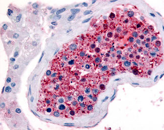
Application
| IHC-P, E |
|---|---|
| Primary Accession | O00144 |
| Reactivity | Human, Mouse, Rabbit, Bovine, Dog |
| Host | Rabbit |
| Clonality | Polyclonal |
| Calculated MW | 64kDa |
| Dilution | IHC-P (5-10 µg/ml) |
| Gene ID | 8326 |
|---|---|
| Other Names | Frizzled-9, Fz-9, hFz9, FzE6, CD349, FZD9, FZD3 |
| Target/Specificity | Human FZD9 / Frizzled 9. BLAST analysis of the peptide immunogen showed no homology with other human proteins, except FZD10 (65%), CARD10 (65%). |
| Reconstitution & Storage | Long term: -70°C; Short term: +4°C |
| Precautions | FZD9 / Frizzled 9 Antibody (N-Terminus) is for research use only and not for use in diagnostic or therapeutic procedures. |
| Name | FZD9 |
|---|---|
| Synonyms | FZD3 |
| Function | Receptor for WNT2 that is coupled to the beta-catenin canonical signaling pathway, which leads to the activation of disheveled proteins, inhibition of GSK-3 kinase, nuclear accumulation of beta-catenin and activation of Wnt target genes (By similarity). Plays a role in neuromuscular junction (NMJ) assembly by negatively regulating the clustering of acetylcholine receptors (AChR) through the beta-catenin canonical signaling pathway (By similarity). May play a role in neural progenitor cells (NPCs) viability through the beta- catenin canonical signaling pathway by negatively regulating cell cycle arrest leading to inhibition of neuron apoptotic process (PubMed:27509850). During hippocampal development, regulates neuroblast proliferation and apoptotic cell death. Controls bone formation through non canonical Wnt signaling mediated via ISG15. Positively regulates bone regeneration through non canonical Wnt signaling (By similarity). |
| Cellular Location | Cell membrane {ECO:0000250|UniProtKB:Q9R216}; Multi-pass membrane protein. Note=Relocalizes DVL1 to the cell membrane leading to phosphorylation of DVL1 and AXIN1 relocalization to the cell membrane. {ECO:0000250|UniProtKB:Q8K4C8} |
| Tissue Location | Expressed predominantly in adult and fetal brain, testis, eye, skeletal muscle and kidney. Moderately expressed in pancreas, thyroid, adrenal cortex, small intestine and stomach Detected in fetal liver and kidney. Expressed in neural progenitor cells (PubMed:27509850). |
| Volume | 50 µl |

Thousands of laboratories across the world have published research that depended on the performance of antibodies from Abcepta to advance their research. Check out links to articles that cite our products in major peer-reviewed journals, organized by research category.
info@abcepta.com, and receive a free "I Love Antibodies" mug.

Provided below are standard protocols that you may find useful for product applications.
Background
Receptor for Wnt proteins. Most of frizzled receptors are coupled to the beta-catenin canonical signaling pathway, which leads to the activation of disheveled proteins, inhibition of GSK- 3 kinase, nuclear accumulation of beta-catenin and activation of Wnt target genes. A second signaling pathway involving PKC and calcium fluxes has been seen for some family members, but it is not yet clear if it represents a distinct pathway or if it can be integrated in the canonical pathway, as PKC seems to be required for Wnt-mediated inactivation of GSK-3 kinase. Both pathways seem to involve interactions with G-proteins. May be involved in transduction and intercellular transmission of polarity information during tissue morphogenesis and/or in differentiated tissues.
References
Wang Y.-K.,et al.Hum. Mol. Genet. 6:465-472(1997).
Hillier L.W.,et al.Nature 424:157-164(2003).
Tanaka S.,et al.Proc. Natl. Acad. Sci. U.S.A. 95:10164-10169(1998).
If you have used an Abcepta product and would like to share how it has performed, please click on the "Submit Review" button and provide the requested information. Our staff will examine and post your review and contact you if needed.
If you have any additional inquiries please email technical services at tech@abcepta.com.
Ordering Information
Other Products
Shipping Information
