


 Foundational characteristics of cancer include proliferation, angiogenesis, migration, evasion of apoptosis, and cellular immortality. Find key markers for these cellular processes and antibodies to detect them.
 The SUMOplot™ Analysis Program predicts and scores sumoylation sites in your protein. SUMOylation is a post-translational modification involved in various cellular processes, such as nuclear-cytosolic transport, transcriptional regulation, apoptosis, protein stability, response to stress, and progression through the cell cycle.
 The Autophagy Receptor Motif Plotter predicts and scores autophagy receptor binding sites in your protein. Identifying proteins connected to this pathway is critical to understanding the role of autophagy in physiological as well as pathological processes such as development, differentiation, neurodegenerative diseases, stress, infection, and cancer.
ACVR1C / ALK7 Antibody (Internal)
Rabbit Polyclonal Antibody
- SPECIFICATION
- CITATIONS
- PROTOCOLS
- BACKGROUND

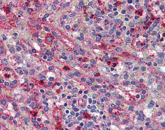
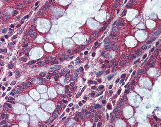
Application
| IHC-P |
|---|---|
| Primary Accession | Q8NER5 |
| Reactivity | Human, Pig, Bovine |
| Host | Rabbit |
| Clonality | Polyclonal |
| Calculated MW | 55kDa |
| Dilution | IHC-P (5 µg/ml) |
| Gene ID | 130399 |
|---|---|
| Other Names | Activin receptor type-1C, 2.7.11.30, Activin receptor type IC, ACTR-IC, Activin receptor-like kinase 7, ALK-7, ACVR1C (HGNC:18123) |
| Target/Specificity | Human ACVR1C. BLAST analysis of the peptide immunogen showed no homology with other human proteins. |
| Reconstitution & Storage | Long term: -70°C; Short term: +4°C |
| Precautions | ACVR1C / ALK7 Antibody (Internal) is for research use only and not for use in diagnostic or therapeutic procedures. |
| Name | ACVR1C (HGNC:18123) |
|---|---|
| Function | Serine/threonine protein kinase which forms a receptor complex on ligand binding. The receptor complex consists of 2 type II and 2 type I transmembrane serine/threonine kinases. Type II receptors phosphorylate and activate type I receptors which autophosphorylate, then bind and activate SMAD transcriptional regulators, SMAD2 and SMAD3. Receptor for activin AB, activin B, activin E and NODAL. Upon NODAL binding, activation results in increased apoptosis and reduced proliferation through suppression of AKT signaling and the activation of Smad2-dependent signaling pathway in pancreatic beta-cells, trophoblasts, epithelial or neuronal cells (PubMed:15531507, PubMed:15150278). Acts as a positive regulator for macrophage activation partially through down-regulation of PPARG expression (By similarity). |
| Cellular Location | Membrane; Single- pass type I membrane protein |
| Tissue Location | Present in pancreas, heart, colon, small intestine, ovary and the hippocampus, medulla oblongata and putamen of the brain Isoform 1, isoform 2, isoform 3 and isoform 4 are all expressed in the placenta throughout pregnancy. |
| Volume | 50 µl |

Thousands of laboratories across the world have published research that depended on the performance of antibodies from Abcepta to advance their research. Check out links to articles that cite our products in major peer-reviewed journals, organized by research category.
info@abcepta.com, and receive a free "I Love Antibodies" mug.

Provided below are standard protocols that you may find useful for product applications.
Background
Serine/threonine protein kinase which forms a receptor complex on ligand binding. The receptor complex consisting of 2 type II and 2 type I transmembrane serine/threonine kinases. Type II receptors phosphorylate and activate type I receptors which autophosphorylate, then bind and activate SMAD transcriptional regulators, SMAD2 and SMAD3. Receptor for activin AB, activin B and NODAL. Plays a role in cell differentiation, growth arrest and apoptosis.
References
Bondestam J.,et al.Cytogenet. Cell Genet. 95:157-162(2001).
Roberts H.J.,et al.Biol. Reprod. 68:1719-1726(2003).
Hillier L.W.,et al.Nature 434:724-731(2005).
Xu G.,et al.J. Clin. Endocrinol. Metab. 89:5523-5534(2004).
Greenman C.,et al.Nature 446:153-158(2007).
If you have used an Abcepta product and would like to share how it has performed, please click on the "Submit Review" button and provide the requested information. Our staff will examine and post your review and contact you if needed.
If you have any additional inquiries please email technical services at tech@abcepta.com.
Ordering Information
Other Products
Shipping Information
