


 Foundational characteristics of cancer include proliferation, angiogenesis, migration, evasion of apoptosis, and cellular immortality. Find key markers for these cellular processes and antibodies to detect them.
 The SUMOplot™ Analysis Program predicts and scores sumoylation sites in your protein. SUMOylation is a post-translational modification involved in various cellular processes, such as nuclear-cytosolic transport, transcriptional regulation, apoptosis, protein stability, response to stress, and progression through the cell cycle.
 The Autophagy Receptor Motif Plotter predicts and scores autophagy receptor binding sites in your protein. Identifying proteins connected to this pathway is critical to understanding the role of autophagy in physiological as well as pathological processes such as development, differentiation, neurodegenerative diseases, stress, infection, and cancer.
NEK9 Antibody (Kinase Domain)
Rabbit Polyclonal Antibody
- SPECIFICATION
- CITATIONS
- PROTOCOLS
- BACKGROUND

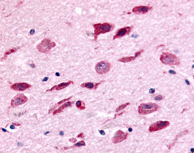
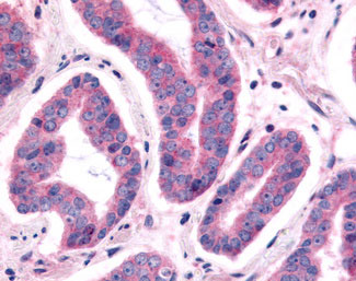
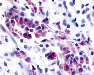
Application
| IHC-P |
|---|---|
| Primary Accession | Q8TD19 |
| Reactivity | Human, Mouse, Rabbit, Monkey, Pig, Chicken, Horse, Bovine |
| Host | Rabbit |
| Clonality | Polyclonal |
| Calculated MW | 107kDa |
| Dilution | IHC-P (4 µg/ml) |
| Gene ID | 91754 |
|---|---|
| Other Names | Serine/threonine-protein kinase Nek9, 2.7.11.1, Nercc1 kinase, Never in mitosis A-related kinase 9, NimA-related protein kinase 9, NimA-related kinase 8, Nek8, NEK9, KIAA1995, NEK8, NERCC |
| Target/Specificity | Human NEK9. BLAST analysis of the peptide immunogen showed no homology with other human proteins. |
| Reconstitution & Storage | Long term: -70°C; Short term: +4°C |
| Precautions | NEK9 Antibody (Kinase Domain) is for research use only and not for use in diagnostic or therapeutic procedures. |
| Name | NEK9 {ECO:0000303|PubMed:12840024, ECO:0000312|HGNC:HGNC:18591} |
|---|---|
| Function | Pleiotropic regulator of mitotic progression, participating in the control of spindle dynamics and chromosome separation (PubMed:12101123, PubMed:12840024, PubMed:14660563, PubMed:19941817). Phosphorylates different histones, myelin basic protein, beta-casein, and BICD2 (PubMed:11864968). Phosphorylates histone H3 on serine and threonine residues and beta-casein on serine residues (PubMed:11864968). Important for G1/S transition and S phase progression (PubMed:12840024, PubMed:14660563, PubMed:19941817). Phosphorylates NEK6 and NEK7 and stimulates their activity by releasing the autoinhibitory functions of Tyr-108 and Tyr-97 respectively (PubMed:12840024, PubMed:14660563, PubMed:19941817, PubMed:26522158). |
| Cellular Location | Cytoplasm. Nucleus |
| Tissue Location | Most abundant in heart, liver, kidney and testis. Also expressed in smooth muscle cells and fibroblasts |

Thousands of laboratories across the world have published research that depended on the performance of antibodies from Abcepta to advance their research. Check out links to articles that cite our products in major peer-reviewed journals, organized by research category.
info@abcepta.com, and receive a free "I Love Antibodies" mug.

Provided below are standard protocols that you may find useful for product applications.
Background
Pleiotropic regulator of mitotic progression, participating in the control of spindle dynamics and chromosome separation. Phosphorylates different histones, myelin basic protein, beta-casein, and BICD2. Phosphorylates histone H3 on serine and threonine residues and beta-casein on serine residues. Important for G1/S transition and S phase progression. Phosphorylates NEK6 and NEK7 and stimulates their activity by releasing the autoinhibitory functions of Tyr-108 and Tyr-97 respectively.
References
Holland P.M.,et al.J. Biol. Chem. 277:16229-16240(2002).
Roig J.,et al.Genes Dev. 16:1640-1658(2002).
Ohara O.,et al.DNA Res. 9:47-57(2002).
Heilig R.,et al.Nature 421:601-607(2003).
Belham C.,et al.J. Biol. Chem. 278:34897-34909(2003).
If you have used an Abcepta product and would like to share how it has performed, please click on the "Submit Review" button and provide the requested information. Our staff will examine and post your review and contact you if needed.
If you have any additional inquiries please email technical services at tech@abcepta.com.
Ordering Information
Other Products
Shipping Information
