


 Foundational characteristics of cancer include proliferation, angiogenesis, migration, evasion of apoptosis, and cellular immortality. Find key markers for these cellular processes and antibodies to detect them.
 The SUMOplot™ Analysis Program predicts and scores sumoylation sites in your protein. SUMOylation is a post-translational modification involved in various cellular processes, such as nuclear-cytosolic transport, transcriptional regulation, apoptosis, protein stability, response to stress, and progression through the cell cycle.
 The Autophagy Receptor Motif Plotter predicts and scores autophagy receptor binding sites in your protein. Identifying proteins connected to this pathway is critical to understanding the role of autophagy in physiological as well as pathological processes such as development, differentiation, neurodegenerative diseases, stress, infection, and cancer.
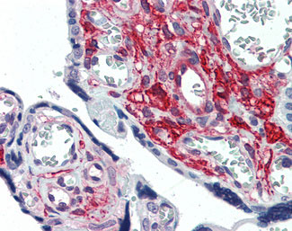
GLG1 / MG160 Antibody (Internal)
Rabbit Polyclonal Antibody
- SPECIFICATION
- CITATIONS
- PROTOCOLS
- BACKGROUND

Application
| IHC-P |
|---|---|
| Primary Accession | Q92896 |
| Reactivity | Human, Mouse, Rabbit, Hamster, Monkey, Pig, Horse, Bovine, Guinea Pig, Dog |
| Host | Rabbit |
| Clonality | Polyclonal |
| Calculated MW | 135kDa |
| Dilution | IHC-P (15 µg/ml) |
| Gene ID | 2734 |
|---|---|
| Other Names | Golgi apparatus protein 1, CFR-1, Cysteine-rich fibroblast growth factor receptor, E-selectin ligand 1, ESL-1, Golgi sialoglycoprotein MG-160, GLG1, CFR1, ESL1, MG160 |
| Target/Specificity | Human GLG1. BLAST analysis of the peptide immunogen showed no homology with other human proteins. |
| Reconstitution & Storage | Long term: -70°C; Short term: +4°C |
| Precautions | GLG1 / MG160 Antibody (Internal) is for research use only and not for use in diagnostic or therapeutic procedures. |
| Name | GLG1 |
|---|---|
| Synonyms | CFR1, ESL1, MG160 |
| Function | Binds fibroblast growth factor and E-selectin (cell-adhesion lectin on endothelial cells mediating the binding of neutrophils). |
| Cellular Location | Golgi apparatus membrane; Single-pass type I membrane protein. Golgi outpost {ECO:0000250|UniProtKB:Q62638}. Cytoplasm, cytoskeleton, microtubule organizing center {ECO:0000250|UniProtKB:Q62638}. Note=Golgi medial cisternae. Localizes to the postsynaptic Golgi apparatus region, also named Golgi outpost, which shapes dendrite morphology by functioning as sites of acentrosomal microtubule nucleation. {ECO:0000250|UniProtKB:Q62638} |
| Tissue Location | Widely expressed. Highest levels in pancreas, skeletal muscle, placenta, heart, testis and ovary. Also found in the kidney, liver, lung and brain. |
| Volume | 50 µl |

Thousands of laboratories across the world have published research that depended on the performance of antibodies from Abcepta to advance their research. Check out links to articles that cite our products in major peer-reviewed journals, organized by research category.
info@abcepta.com, and receive a free "I Love Antibodies" mug.

Provided below are standard protocols that you may find useful for product applications.
Background
Binds fibroblast growth factor and E-selectin (cell- adhesion lectin on endothelial cells mediating the binding of neutrophils).
References
Mourelatos Z.,et al.DNA Cell Biol. 15:1121-1128(1996).
Wu M.,et al.Submitted (JUN-1995) to the EMBL/GenBank/DDBJ databases.
Ota T.,et al.Nat. Genet. 36:40-45(2004).
Martin J.,et al.Nature 432:988-994(2004).
Mural R.J.,et al.Submitted (SEP-2005) to the EMBL/GenBank/DDBJ databases.
If you have used an Abcepta product and would like to share how it has performed, please click on the "Submit Review" button and provide the requested information. Our staff will examine and post your review and contact you if needed.
If you have any additional inquiries please email technical services at tech@abcepta.com.
Ordering Information
Other Products
Shipping Information
