


 Foundational characteristics of cancer include proliferation, angiogenesis, migration, evasion of apoptosis, and cellular immortality. Find key markers for these cellular processes and antibodies to detect them.
 The SUMOplot™ Analysis Program predicts and scores sumoylation sites in your protein. SUMOylation is a post-translational modification involved in various cellular processes, such as nuclear-cytosolic transport, transcriptional regulation, apoptosis, protein stability, response to stress, and progression through the cell cycle.
 The Autophagy Receptor Motif Plotter predicts and scores autophagy receptor binding sites in your protein. Identifying proteins connected to this pathway is critical to understanding the role of autophagy in physiological as well as pathological processes such as development, differentiation, neurodegenerative diseases, stress, infection, and cancer.
> home > Products > Primary Antibodies > Antibody Collections > Catalog Updated > GLG1 Antibody (C-term)
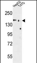
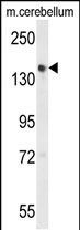
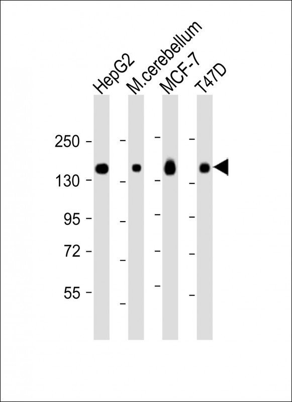
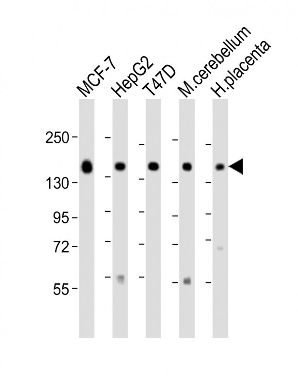
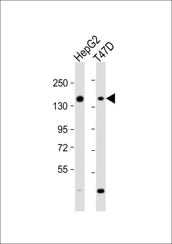
GLG1 Antibody (C-term)
Affinity Purified Rabbit Polyclonal Antibody (Pab)
- SPECIFICATION
- CITATIONS: 1
- PROTOCOLS
- BACKGROUND

Application
| WB, IHC-P, E |
|---|---|
| Primary Accession | Q92896 |
| Other Accession | Q62638, Q61543, Q9Z1E9, Q02391 |
| Reactivity | Human, Mouse |
| Predicted | Chicken, Hamster, Rat |
| Host | Rabbit |
| Clonality | Polyclonal |
| Isotype | Rabbit IgG |
| Calculated MW | 134552 Da |
| Antigen Region | 1152-1179 aa |
| Gene ID | 2734 |
|---|---|
| Other Names | Golgi apparatus protein 1, CFR-1, Cysteine-rich fibroblast growth factor receptor, E-selectin ligand 1, ESL-1, Golgi sialoglycoprotein MG-160, GLG1, CFR1, ESL1, MG160 |
| Target/Specificity | This GLG1 antibody is generated from rabbits immunized with a KLH conjugated synthetic peptide between 1152-1179 amino acids from the C-terminal region of human GLG1. |
| Dilution | WB~~1:2000 IHC-P~~1:50~100 E~~Use at an assay dependent concentration. |
| Format | Purified polyclonal antibody supplied in PBS with 0.09% (W/V) sodium azide. This antibody is purified through a protein A column, followed by peptide affinity purification. |
| Storage | Maintain refrigerated at 2-8°C for up to 2 weeks. For long term storage store at -20°C in small aliquots to prevent freeze-thaw cycles. |
| Precautions | GLG1 Antibody (C-term) is for research use only and not for use in diagnostic or therapeutic procedures. |
| Name | GLG1 |
|---|---|
| Synonyms | CFR1, ESL1, MG160 |
| Function | Binds fibroblast growth factor and E-selectin (cell-adhesion lectin on endothelial cells mediating the binding of neutrophils). |
| Cellular Location | Golgi apparatus membrane; Single-pass type I membrane protein. Golgi outpost {ECO:0000250|UniProtKB:Q62638}. Cytoplasm, cytoskeleton, microtubule organizing center {ECO:0000250|UniProtKB:Q62638}. Note=Golgi medial cisternae. Localizes to the postsynaptic Golgi apparatus region, also named Golgi outpost, which shapes dendrite morphology by functioning as sites of acentrosomal microtubule nucleation. {ECO:0000250|UniProtKB:Q62638} |
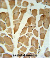
| Tissue Location | Widely expressed. Highest levels in pancreas, skeletal muscle, placenta, heart, testis and ovary. Also found in the kidney, liver, lung and brain. |
Research Areas
Citations ( 0 )

Application Protocols
Provided below are standard protocols that you may find useful for product applications.
References
Dastani, Z., et al. Eur. J. Hum. Genet. 18(3):342-347(2010)
Kibriya, M.G., et al. Breast Cancer Res. Treat. 114(3):463-477(2009)
Antoine, M., et al. Oncol. Rep. 21(2):357-362(2009)
Ahn, J., et al. J. Cell. Sci. 118 (PT 8), 1725-1731 (2005)
Bouwmeester, T., et al. Nat. Cell Biol. 6(2):97-105(2004)
Abcepta welcomes feedback from its customers.
If you have used an Abcepta product and would like to share how it has performed, please click on the "Submit Review" button and provide the requested information. Our staff will examine and post your review and contact you if needed.
If you have any additional inquiries please email technical services at tech@abcepta.com.
$ 150.00
$ 385.00
Cat# AP9839b
Ordering Information
United States
AlbaniaAustraliaAustriaBelgiumBosnia & HerzegovinaBrazilBulgariaCanadaCentral AmericaChinaCroatiaCyprusCzech RepublicDenmarkEstoniaFinlandFranceGermanyGreeceHong KongHungaryIcelandIndiaIndonesiaIrelandIsraelItalyJapanLatviaLithuaniaLuxembourgMacedoniaMalaysiaMaltaMexicoNetherlandsNew ZealandNorwayPakistanPolandPortugalRomaniaSerbiaSingaporeSlovakiaSloveniaSouth AfricaSouth KoreaSpainSwedenSwitzerlandTaiwanTurkeyUnited KingdomUnited StatesVietnamWorldwideOthers
USA Headquarters
(888) 735-7227 / (858) 622-0099 or (858) 875-1900
Other Products
Shipping Information
Domestic orders (in stock items)
Shipped out the same day. Orders placed after 1 PM (PST) will ship out the next business day.
International orders
Contact your local distributors
