


 Foundational characteristics of cancer include proliferation, angiogenesis, migration, evasion of apoptosis, and cellular immortality. Find key markers for these cellular processes and antibodies to detect them.
 The SUMOplot™ Analysis Program predicts and scores sumoylation sites in your protein. SUMOylation is a post-translational modification involved in various cellular processes, such as nuclear-cytosolic transport, transcriptional regulation, apoptosis, protein stability, response to stress, and progression through the cell cycle.
 The Autophagy Receptor Motif Plotter predicts and scores autophagy receptor binding sites in your protein. Identifying proteins connected to this pathway is critical to understanding the role of autophagy in physiological as well as pathological processes such as development, differentiation, neurodegenerative diseases, stress, infection, and cancer.
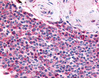
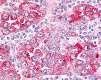
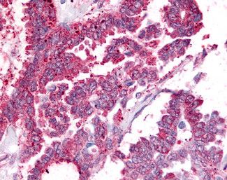
PGES / PTGES Antibody (Cytoplasmic Domain)
Rabbit Polyclonal Antibody
- SPECIFICATION
- CITATIONS
- PROTOCOLS
- BACKGROUND

Application
| IHC-P |
|---|---|
| Primary Accession | O14684 |
| Reactivity | Human, Rabbit |
| Host | Rabbit |
| Clonality | Polyclonal |
| Calculated MW | 17kDa |
| Dilution | IHC-P (5-10 µg/ml) |
| Gene ID | 9536 |
|---|---|
| Other Names | Prostaglandin E synthase, 5.3.99.3, Microsomal glutathione S-transferase 1-like 1, MGST1-L1, Microsomal prostaglandin E synthase 1, MPGES-1, p53-induced gene 12 protein, PTGES, MGST1L1, MPGES1, PGES, PIG12 |
| Target/Specificity | Human PTGES. BLAST analysis of the peptide immunogen showed no homology with other human proteins. |
| Reconstitution & Storage | Long term: -70°C; Short term: +4°C |
| Precautions | PGES / PTGES Antibody (Cytoplasmic Domain) is for research use only and not for use in diagnostic or therapeutic procedures. |
| Name | PTGES |
|---|---|
| Function | Terminal enzyme of the cyclooxygenase (COX)-2-mediated prostaglandin E2 (PGE2) biosynthetic pathway. Catalyzes the glutathione-dependent oxidoreduction of prostaglandin endoperoxide H2 (PGH2) to prostaglandin E2 (PGE2) in response to inflammatory stimuli (PubMed:10377395, PubMed:10869354, PubMed:12244105, PubMed:12460774, PubMed:12672824, PubMed:18682561). Plays a key role in inflammation response, fever and pain (By similarity). Also catalyzes the oxidoreduction of endocannabinoids into prostaglandin glycerol esters and PGG2 into 15-hydroperoxy-PGE2 (PubMed:12244105, PubMed:12672824). In addition, displays low glutathione transferase and glutathione- dependent peroxidase activities, toward 1-chloro-2,4-dinitrobenzene and 5-hydroperoxyicosatetraenoic acid (5-HPETE), respectively (PubMed:12672824). |
| Cellular Location | Membrane; Multi-pass membrane protein. Cytoplasm, perinuclear region. Note=Colocalizes with PTGS1/COX-1 and PTGS2/COX-2 in the perinuclear compartment |
| Volume | 100 µl |

Thousands of laboratories across the world have published research that depended on the performance of antibodies from Abcepta to advance their research. Check out links to articles that cite our products in major peer-reviewed journals, organized by research category.
info@abcepta.com, and receive a free "I Love Antibodies" mug.

Provided below are standard protocols that you may find useful for product applications.
Background
Catalyzes the oxidoreduction of prostaglandin endoperoxide H2 (PGH2) to prostaglandin E2 (PGE2).
References
Polyak K.,et al.Nature 389:300-306(1997).
Jakobsson P.-J.,et al.Proc. Natl. Acad. Sci. U.S.A. 96:7220-7225(1999).
Forsberg L.,et al.FEBS Lett. 471:78-82(2000).
Ota T.,et al.Nat. Genet. 36:40-45(2004).
Humphray S.J.,et al.Nature 429:369-374(2004).
If you have used an Abcepta product and would like to share how it has performed, please click on the "Submit Review" button and provide the requested information. Our staff will examine and post your review and contact you if needed.
If you have any additional inquiries please email technical services at tech@abcepta.com.
Ordering Information
Other Products
Shipping Information
