


 Foundational characteristics of cancer include proliferation, angiogenesis, migration, evasion of apoptosis, and cellular immortality. Find key markers for these cellular processes and antibodies to detect them.
 The SUMOplot™ Analysis Program predicts and scores sumoylation sites in your protein. SUMOylation is a post-translational modification involved in various cellular processes, such as nuclear-cytosolic transport, transcriptional regulation, apoptosis, protein stability, response to stress, and progression through the cell cycle.
 The Autophagy Receptor Motif Plotter predicts and scores autophagy receptor binding sites in your protein. Identifying proteins connected to this pathway is critical to understanding the role of autophagy in physiological as well as pathological processes such as development, differentiation, neurodegenerative diseases, stress, infection, and cancer.
GCNT3 Antibody (N-Terminus)
Rabbit Polyclonal Antibody
- SPECIFICATION
- CITATIONS
- PROTOCOLS
- BACKGROUND

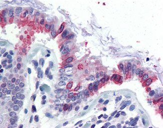
Application
| IHC-P |
|---|---|
| Primary Accession | O95395 |
| Reactivity | Human |
| Host | Rabbit |
| Clonality | Polyclonal |
| Calculated MW | 51kDa |
| Dilution | IHC-P (2.5 µg/ml) |
| Gene ID | 9245 |
|---|---|
| Other Names | Beta-1, 3-galactosyl-O-glycosyl-glycoprotein beta-1, 6-N-acetylglucosaminyltransferase 3, 2.4.1.102, 2.4.1.150, C2GnT-mucin type, C2GnT-M, hC2GnT-M, Core 2/core 4 beta-1, 6-N-acetylglucosaminyltransferase, C2/4GnT, GCNT3 |
| Target/Specificity | Human GCNT3. BLAST analysis of the peptide immunogen showed no homology with other human proteins, except NEK8 (60%). |
| Reconstitution & Storage | Long term: -70°C; Short term: +4°C |
| Precautions | GCNT3 Antibody (N-Terminus) is for research use only and not for use in diagnostic or therapeutic procedures. |
| Name | GCNT3 |
|---|---|
| Function | Glycosyltransferase that can synthesize all known mucin beta 6 N-acetylglucosaminides. Mediates core 2 and core 4 O-glycan branching, 2 important steps in mucin-type biosynthesis. Also has I- branching enzyme activity by converting linear into branched poly-N- acetyllactosaminoglycans, leading to introduce the blood group I antigen during embryonic development. |
| Cellular Location | Golgi apparatus membrane; Single- pass type II membrane protein |
| Tissue Location | Primarily expressed in mucus-secreting tissues. Expressed in colon, kidney, small intestine, trachea, and stomach, where mucin is produced. |
| Volume | 50 µl |

Thousands of laboratories across the world have published research that depended on the performance of antibodies from Abcepta to advance their research. Check out links to articles that cite our products in major peer-reviewed journals, organized by research category.
info@abcepta.com, and receive a free "I Love Antibodies" mug.

Provided below are standard protocols that you may find useful for product applications.
Background
Glycosyltransferase that can synthesize all known mucin beta 6 N-acetylglucosaminides. Mediates core 2 and core 4 O-glycan branching, 2 important steps in mucin-type biosynthesis. Has also I-branching enzyme activity by converting linear into branched poly-N-acetyllactosaminoglycans, leading to introduce the blood group I antigen during embryonic development.
References
Yeh J.-C.,et al.J. Biol. Chem. 274:3215-3221(1999).
Schwientek T.,et al.J. Biol. Chem. 274:4504-4512(1999).
Tan S.,et al.Am. J. Respir. Cell Mol. Biol. 36:737-745(2007).
Ishibashi Y.,et al.Glycoconj. J. 22:53-62(2005).
Huang M.-C.,et al.Oncogene 25:3267-3276(2006).
If you have used an Abcepta product and would like to share how it has performed, please click on the "Submit Review" button and provide the requested information. Our staff will examine and post your review and contact you if needed.
If you have any additional inquiries please email technical services at tech@abcepta.com.
Ordering Information
Other Products
Shipping Information
