


 Foundational characteristics of cancer include proliferation, angiogenesis, migration, evasion of apoptosis, and cellular immortality. Find key markers for these cellular processes and antibodies to detect them.
 The SUMOplot™ Analysis Program predicts and scores sumoylation sites in your protein. SUMOylation is a post-translational modification involved in various cellular processes, such as nuclear-cytosolic transport, transcriptional regulation, apoptosis, protein stability, response to stress, and progression through the cell cycle.
 The Autophagy Receptor Motif Plotter predicts and scores autophagy receptor binding sites in your protein. Identifying proteins connected to this pathway is critical to understanding the role of autophagy in physiological as well as pathological processes such as development, differentiation, neurodegenerative diseases, stress, infection, and cancer.
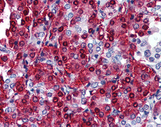
CBLC Antibody (aa444-458)
Rabbit Polyclonal Antibody
- SPECIFICATION
- CITATIONS
- PROTOCOLS
- BACKGROUND

Application
| WB, IHC-P, IF, E, IP |
|---|---|
| Primary Accession | Q9ULV8 |
| Reactivity | Human |
| Host | Rabbit |
| Clonality | Polyclonal |
| Calculated MW | 52kDa |
| Dilution | ELISA (1:10000-1:50000), IHC-P (5 µg/ml), WB (1:500-1:3000) |
| Gene ID | 23624 |
|---|---|
| Other Names | E3 ubiquitin-protein ligase CBL-C, 6.3.2.-, RING finger protein 57, SH3-binding protein CBL-3, SH3-binding protein CBL-C, Signal transduction protein CBL-C, CBLC, CBL3, RNF57 |
| Target/Specificity | Amino acids 444-458 of Human Cbl-c. |
| Reconstitution & Storage | +4°C or -20°C, Avoid repeated freezing and thawing. |
| Precautions | CBLC Antibody (aa444-458) is for research use only and not for use in diagnostic or therapeutic procedures. |
| Name | CBLC |
|---|---|
| Synonyms | CBL3, RNF57 |
| Function | Acts as an E3 ubiquitin-protein ligase, which accepts ubiquitin from specific E2 ubiquitin-conjugating enzymes, and then transfers it to substrates promoting their degradation by the proteasome. Functionally coupled with the E2 ubiquitin-protein ligases UB2D1, UB2D2 and UB2D3. Regulator of EGFR mediated signal transduction; upon EGF activation, ubiquitinates EGFR. Isoform 1, but not isoform 2, inhibits EGF stimulated MAPK1 activation. Promotes ubiquitination of SRC phosphorylated at 'Tyr-419'. In collaboration with CD2AP may act as regulatory checkpoint for Ret signaling by modulating the rate of RET degradation after ligand activation; CD2AP converts it from an inhibitor to a promoter of RET degradation; the function limits the potency of GDNF on neuronal survival. |
| Tissue Location | Ubiquitous.. |
| Volume | 100 µl |

Thousands of laboratories across the world have published research that depended on the performance of antibodies from Abcepta to advance their research. Check out links to articles that cite our products in major peer-reviewed journals, organized by research category.
info@abcepta.com, and receive a free "I Love Antibodies" mug.

Provided below are standard protocols that you may find useful for product applications.
Background
Acts as an E3 ubiquitin-protein ligase, which accepts ubiquitin from specific E2 ubiquitin-conjugating enzymes, and then transfers it to substrates promoting their degradation by the proteasome. Functionally coupled with the E2 ubiquitin-protein ligases UB2D1, UB2D2 and UB2D3. Regulator of EGFR mediated signal transduction; upon EGF activation, ubiquitinates EGFR. Isoform 1, but not isoform 2, inhibits EGF stimulated MAPK1 activation. Promotes ubiquitination of SRC phosphorylated at 'Tyr-419'. In collaboration with CD2AP may act as regulatory checkpoint for Ret signaling by modulating the rate of RET degradation after ligand activation; CD2AP converts it from an inhibitor to a promoter of RET degradation; the function limits the potency of GDNF on neuronal survival.
References
Kim M.,et al.Gene 239:145-154(1999).
Keane M.M.,et al.Oncogene 18:3365-3375(1999).
Kim M.,et al.Oncogene 23:1645-1655(2004).
Tsui C.C.,et al.J. Neurosci. 28:8789-8800(2008).
Ryan P.E.,et al.J. Biol. Chem. 285:23687-23698(2010).
If you have used an Abcepta product and would like to share how it has performed, please click on the "Submit Review" button and provide the requested information. Our staff will examine and post your review and contact you if needed.
If you have any additional inquiries please email technical services at tech@abcepta.com.

Ordering Information
Other Products
Shipping Information
