


 Foundational characteristics of cancer include proliferation, angiogenesis, migration, evasion of apoptosis, and cellular immortality. Find key markers for these cellular processes and antibodies to detect them.
 The SUMOplot™ Analysis Program predicts and scores sumoylation sites in your protein. SUMOylation is a post-translational modification involved in various cellular processes, such as nuclear-cytosolic transport, transcriptional regulation, apoptosis, protein stability, response to stress, and progression through the cell cycle.
 The Autophagy Receptor Motif Plotter predicts and scores autophagy receptor binding sites in your protein. Identifying proteins connected to this pathway is critical to understanding the role of autophagy in physiological as well as pathological processes such as development, differentiation, neurodegenerative diseases, stress, infection, and cancer.
ICAM-1 / CD54 Antibody (clone 15.2)
Mouse Monoclonal Antibody
- SPECIFICATION
- CITATIONS
- PROTOCOLS
- BACKGROUND

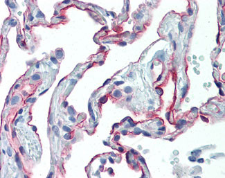
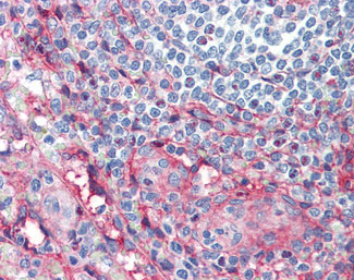
Application
| IHC-P, IP, IHC-F, FC |
|---|---|
| Primary Accession | P05362 |
| Reactivity | Human, Pig |
| Host | Mouse |
| Clonality | Monoclonal |
| Clone Names | 15.2 |
| Calculated MW | 58kDa |
| Dilution | Flo (1:50-1:100), IHC-P (10 µg/ml), |
| Gene ID | 3383 |
|---|---|
| Other Names | Intercellular adhesion molecule 1, ICAM-1, Major group rhinovirus receptor, CD54, ICAM1 |
| Target/Specificity | Recognizes the human CD54 cell surface antigen, a 90kD glycoprotein also known as ICAM-1. CD54 is expressed by many cells following activation by inflammatory mediators. Clone 15.2 is reported to block CD54 function. |
| Reconstitution & Storage | +4°C or -20°C, Avoid repeated freezing and thawing. |
| Precautions | ICAM-1 / CD54 Antibody (clone 15.2) is for research use only and not for use in diagnostic or therapeutic procedures. |
| Name | ICAM1 |
|---|---|
| Function | ICAM proteins are ligands for the leukocyte adhesion protein LFA-1 (integrin alpha-L/beta-2). During leukocyte trans-endothelial migration, ICAM1 engagement promotes the assembly of endothelial apical cups through ARHGEF26/SGEF and RHOG activation. |
| Cellular Location | Membrane; Single-pass type I membrane protein. |

Thousands of laboratories across the world have published research that depended on the performance of antibodies from Abcepta to advance their research. Check out links to articles that cite our products in major peer-reviewed journals, organized by research category.
info@abcepta.com, and receive a free "I Love Antibodies" mug.

Provided below are standard protocols that you may find useful for product applications.
Background
ICAM proteins are ligands for the leukocyte adhesion protein LFA-1 (integrin alpha-L/beta-2). During leukocyte trans- endothelial migration, ICAM1 engagement promotes the assembly of endothelial apical cups through ARHGEF26/SGEF and RHOG activation. In case of rhinovirus infection acts as a cellular receptor for the virus.
References
Simmons D.,et al.Nature 331:624-627(1988).
Staunton D.E.,et al.Cell 52:925-933(1988).
Tomassini J.E.,et al.Proc. Natl. Acad. Sci. U.S.A. 86:4907-4911(1989).
Voraberger G.F.,et al.J. Immunol. 147:2777-2786(1991).
Kalnine N.,et al.Submitted (MAY-2003) to the EMBL/GenBank/DDBJ databases.
If you have used an Abcepta product and would like to share how it has performed, please click on the "Submit Review" button and provide the requested information. Our staff will examine and post your review and contact you if needed.
If you have any additional inquiries please email technical services at tech@abcepta.com.
Ordering Information
Other Products
Shipping Information
