


 Foundational characteristics of cancer include proliferation, angiogenesis, migration, evasion of apoptosis, and cellular immortality. Find key markers for these cellular processes and antibodies to detect them.
 The SUMOplot™ Analysis Program predicts and scores sumoylation sites in your protein. SUMOylation is a post-translational modification involved in various cellular processes, such as nuclear-cytosolic transport, transcriptional regulation, apoptosis, protein stability, response to stress, and progression through the cell cycle.
 The Autophagy Receptor Motif Plotter predicts and scores autophagy receptor binding sites in your protein. Identifying proteins connected to this pathway is critical to understanding the role of autophagy in physiological as well as pathological processes such as development, differentiation, neurodegenerative diseases, stress, infection, and cancer.
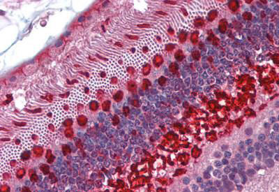
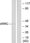
ARR3 / Cone Arrestin Antibody (aa339-388)
Rabbit Polyclonal Antibody
- SPECIFICATION
- CITATIONS
- PROTOCOLS
- BACKGROUND

Application
| WB, IHC, E |
|---|---|
| Primary Accession | P36575 |
| Reactivity | Human |
| Host | Rabbit |
| Clonality | Polyclonal |
| Calculated MW | 43kDa |
| Dilution | ELISA (1:5000), IHC (20 µg/ml), WB (1:500-1:1000) |
| Gene ID | 407 |
|---|---|
| Other Names | Arrestin-C, Cone arrestin, C-arrestin, cArr, Retinal cone arrestin-3, X-arrestin, ARR3, ARRX, CAR |
| Target/Specificity | ARR3 Antibody detects endogenous levels of total ARR3 protein. |
| Reconstitution & Storage | Store at -20°C for up to one year. |
| Precautions | ARR3 / Cone Arrestin Antibody (aa339-388) is for research use only and not for use in diagnostic or therapeutic procedures. |
| Name | ARR3 |
|---|---|
| Synonyms | ARRX, CAR |
| Function | May play a role in an as yet undefined retina-specific signal transduction. Could bind to photoactivated-phosphorylated red/green opsins. |
| Cellular Location | Photoreceptor inner segment {ECO:0000250|UniProtKB:Q9EQP6}. Cell projection, cilium, photoreceptor outer segment {ECO:0000250|UniProtKB:Q9EQP6} |
| Tissue Location | Inner and outer segments, and the inner plexiform regions of the retina |
| Volume | 50 µl |

Thousands of laboratories across the world have published research that depended on the performance of antibodies from Abcepta to advance their research. Check out links to articles that cite our products in major peer-reviewed journals, organized by research category.
info@abcepta.com, and receive a free "I Love Antibodies" mug.

Provided below are standard protocols that you may find useful for product applications.
Background
May play a role in an as yet undefined retina-specific signal transduction. Could binds to photoactivated-phosphorylated red/green opsins.
References
Murakami A.,et al.FEBS Lett. 334:203-209(1993).
Craft C.M.,et al.J. Biol. Chem. 269:4613-4619(1994).
Craft C.M.,et al.Submitted (NOV-1998) to the EMBL/GenBank/DDBJ databases.
Sakuma H.,et al.FEBS Lett. 382:105-110(1996).
Sakuma H.,et al.Gene 224:87-95(1998).
If you have used an Abcepta product and would like to share how it has performed, please click on the "Submit Review" button and provide the requested information. Our staff will examine and post your review and contact you if needed.
If you have any additional inquiries please email technical services at tech@abcepta.com.
Ordering Information
Other Products
Shipping Information
