


 Foundational characteristics of cancer include proliferation, angiogenesis, migration, evasion of apoptosis, and cellular immortality. Find key markers for these cellular processes and antibodies to detect them.
 The SUMOplot™ Analysis Program predicts and scores sumoylation sites in your protein. SUMOylation is a post-translational modification involved in various cellular processes, such as nuclear-cytosolic transport, transcriptional regulation, apoptosis, protein stability, response to stress, and progression through the cell cycle.
 The Autophagy Receptor Motif Plotter predicts and scores autophagy receptor binding sites in your protein. Identifying proteins connected to this pathway is critical to understanding the role of autophagy in physiological as well as pathological processes such as development, differentiation, neurodegenerative diseases, stress, infection, and cancer.
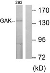
GAK Antibody (aa101-150)
Rabbit Polyclonal Antibody
- SPECIFICATION
- CITATIONS
- PROTOCOLS
- BACKGROUND

Application
| WB, IHC-P, E |
|---|---|
| Primary Accession | O14976 |
| Reactivity | Human, Mouse |
| Host | Rabbit |
| Clonality | Polyclonal |
| Calculated MW | 143kDa |
| Dilution | ELISA (1:5000), IHC-P (20 µg/ml), WB (1:500-1:1000) |
| Gene ID | 2580 |
|---|---|
| Other Names | Cyclin-G-associated kinase, 2.7.11.1, GAK |
| Target/Specificity | GAK Antibody detects endogenous levels of total GAK protein. |
| Reconstitution & Storage | Store at -20°C for up to one year. |
| Precautions | GAK Antibody (aa101-150) is for research use only and not for use in diagnostic or therapeutic procedures. |
| Name | GAK (HGNC:4113) |
|---|---|
| Function | Associates with cyclin G and CDK5. Seems to act as an auxilin homolog that is involved in the uncoating of clathrin-coated vesicles by Hsc70 in non-neuronal cells. Expression oscillates slightly during the cell cycle, peaking at G1 (PubMed:10625686). May play a role in clathrin-mediated endocytosis and intracellular trafficking, and in the dynamics of clathrin assembly/disassembly (PubMed:18489706). |
| Cellular Location | Cytoplasm, perinuclear region. Golgi apparatus, trans-Golgi network. Cell junction, focal adhesion. Cytoplasmic vesicle, clathrin-coated vesicle. Note=Localizes to the perinuclear area and to the trans-Golgi network. Also seen on the plasma membrane, probably at focal adhesions. Recruitment to clathrin- coated vesicles depends on temporal variations in phosphoinositide composition of clathrin-coated vesicles (PubMed:31962345) |
| Tissue Location | Ubiquitous. Highest in testis. |
| Volume | 50 µl |

Thousands of laboratories across the world have published research that depended on the performance of antibodies from Abcepta to advance their research. Check out links to articles that cite our products in major peer-reviewed journals, organized by research category.
info@abcepta.com, and receive a free "I Love Antibodies" mug.

Provided below are standard protocols that you may find useful for product applications.
Background
Associates with cyclin G and CDK5. Seems to act as an auxilin homolog that is involved in the uncoating of clathrin- coated vesicles by Hsc70 in non-neuronal cells. Expression oscillates slightly during the cell cycle, peaking at G1.
References
Kimura S.H.,et al.Genomics 44:179-187(1997).
Greener T.,et al.J. Biol. Chem. 275:1365-1370(2000).
Daub H.,et al.Mol. Cell 31:438-448(2008).
Dephoure N.,et al.Proc. Natl. Acad. Sci. U.S.A. 105:10762-10767(2008).
Gauci S.,et al.Anal. Chem. 81:4493-4501(2009).
If you have used an Abcepta product and would like to share how it has performed, please click on the "Submit Review" button and provide the requested information. Our staff will examine and post your review and contact you if needed.
If you have any additional inquiries please email technical services at tech@abcepta.com.
Ordering Information
Other Products
Shipping Information
