


 Foundational characteristics of cancer include proliferation, angiogenesis, migration, evasion of apoptosis, and cellular immortality. Find key markers for these cellular processes and antibodies to detect them.
 The SUMOplot™ Analysis Program predicts and scores sumoylation sites in your protein. SUMOylation is a post-translational modification involved in various cellular processes, such as nuclear-cytosolic transport, transcriptional regulation, apoptosis, protein stability, response to stress, and progression through the cell cycle.
 The Autophagy Receptor Motif Plotter predicts and scores autophagy receptor binding sites in your protein. Identifying proteins connected to this pathway is critical to understanding the role of autophagy in physiological as well as pathological processes such as development, differentiation, neurodegenerative diseases, stress, infection, and cancer.
P2RX7 / P2X7 Antibody (C-Terminus)
Rabbit Polyclonal Antibody
- SPECIFICATION
- CITATIONS
- PROTOCOLS
- BACKGROUND

Application
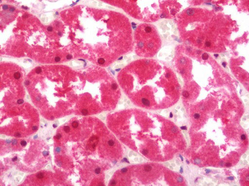
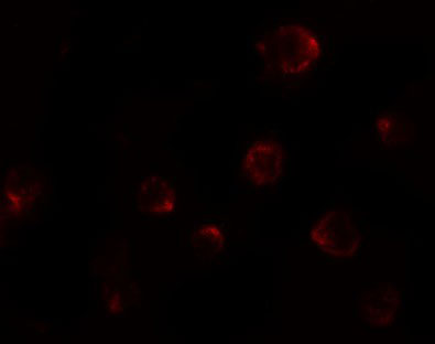
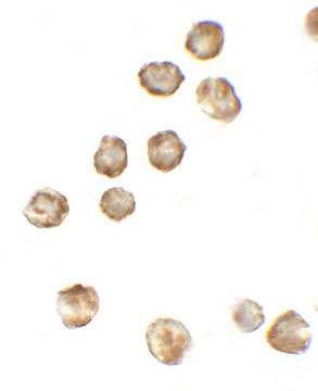
| WB, IHC-P, IF, ICC, E |
|---|---|
| Primary Accession | Q99572 |
| Reactivity | Human, Mouse |
| Host | Rabbit |
| Clonality | Polyclonal |
| Calculated MW | 69kDa |
| Dilution | ICC (5 µg/ml), IF (20 µg/ml), IHC-P (5 µg/ml), WB (1-2 µg/ml) , |
| Gene ID | 5027 |
|---|---|
| Other Names | P2X purinoceptor 7, P2X7, ATP receptor, P2Z receptor, Purinergic receptor, P2RX7 |
| Target/Specificity | P2RX7 antibody is human, mouse, and rat reactive. Multiple isoforms of P2RX7 are known to exist. |
| Reconstitution & Storage | Long term: -20°C; Short term: +4°C. Avoid repeat freeze-thaw cycles. |
| Precautions | P2RX7 / P2X7 Antibody (C-Terminus) is for research use only and not for use in diagnostic or therapeutic procedures. |
| Name | P2RX7 |
|---|---|
| Function | ATP-gated nonselective transmembrane cation channel that requires high millimolar concentrations of ATP for activation (PubMed:17483156, PubMed:25281740, PubMed:9038151). Upon ATP binding, it rapidly opens to allow the influx of small cations Na(+) and Ca(2+), and the K(+) efflux (PubMed:17483156, PubMed:20453110, PubMed:28235784, PubMed:39262850). Also has the ability to form a large pore in the cell membrane, allowing the passage of large cationic molecules (PubMed:17483156). In microglia, may mediate NADPH transport across the plasma membrane (PubMed:39142135). In immune cells, P2RX7 acts as a molecular sensor in pathological inflammatory states by detecting and responding to high local concentrations of extracellar ATP. In microglial cells, P2RX7 activation leads to the release of pro- inflammatory cytokines, such as IL-1beta and IL-18, through the activation of the NLRP3 inflammasome and caspase-1 (PubMed:26877061). Cooperates with KCNK6 to activate NLRP3 inflammasome (By similarity). Activates death pathways leading to apoptosis and autophagy (PubMed:21821797, PubMed:23303206, PubMed:28326637). Activates death pathways leading to pyroptosis (By similarity). |
| Cellular Location | Cell membrane; Multi-pass membrane protein {ECO:0000250|UniProtKB:Q64663} |
| Tissue Location | Widely expressed with highest levels in brain and immune tissues. |

Thousands of laboratories across the world have published research that depended on the performance of antibodies from Abcepta to advance their research. Check out links to articles that cite our products in major peer-reviewed journals, organized by research category.
info@abcepta.com, and receive a free "I Love Antibodies" mug.

Provided below are standard protocols that you may find useful for product applications.
Background
Receptor for ATP that acts as a ligand-gated ion channel. Responsible for ATP-dependent lysis of macrophages through the formation of membrane pores permeable to large molecules. Could function in both fast synaptic transmission and the ATP-mediated lysis of antigen-presenting cells.
References
Rassendren F.,et al.J. Biol. Chem. 272:5482-5486(1997).
Buell G.N.,et al.Recept. Channels 5:347-354(1998).
Cheewatrakoolpong B.,et al.Biochem. Biophys. Res. Commun. 332:17-27(2005).
Ota T.,et al.Nat. Genet. 36:40-45(2004).
Scherer S.E.,et al.Nature 440:346-351(2006).
If you have used an Abcepta product and would like to share how it has performed, please click on the "Submit Review" button and provide the requested information. Our staff will examine and post your review and contact you if needed.
If you have any additional inquiries please email technical services at tech@abcepta.com.
Ordering Information
Other Products
Shipping Information
