


 Foundational characteristics of cancer include proliferation, angiogenesis, migration, evasion of apoptosis, and cellular immortality. Find key markers for these cellular processes and antibodies to detect them.
 The SUMOplot™ Analysis Program predicts and scores sumoylation sites in your protein. SUMOylation is a post-translational modification involved in various cellular processes, such as nuclear-cytosolic transport, transcriptional regulation, apoptosis, protein stability, response to stress, and progression through the cell cycle.
 The Autophagy Receptor Motif Plotter predicts and scores autophagy receptor binding sites in your protein. Identifying proteins connected to this pathway is critical to understanding the role of autophagy in physiological as well as pathological processes such as development, differentiation, neurodegenerative diseases, stress, infection, and cancer.
FOLH1 / PSMA Antibody (clone 3H5)
Mouse Monoclonal Antibody
- SPECIFICATION
- CITATIONS
- PROTOCOLS
- BACKGROUND

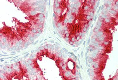
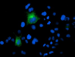
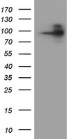
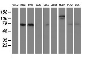
Application
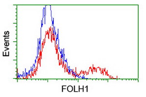
| WB, IHC-P, IF, FC |
|---|---|
| Primary Accession | Q04609 |
| Other Accession | 2346 |
| Reactivity | Human, Mouse |
| Host | Mouse |
| Clonality | Monoclonal |
| Isotype | IgG1 |
| Clone Names | 3H5 |
| Calculated MW | 84331 Da |
| Dilution | Flo (1:100), IF (1:100), IHC-P (1:150), WB (1:500 - 1:1000), |
| Gene ID | 2346 |
|---|---|
| Other Names | FOLH1, Folate hydrolase 1, GCPII, Glutamate carboxypeptidase II, Folate hydrolase, GCP2, NAALAD1, NAALAdase, NAALADase I, PSM, FGCP, FOLH, Glutamate carboxylase II, Glutamate carboxypeptidase 2, MGCP, PSMA |
| Target/Specificity | Human PSMA |
| Reconstitution & Storage | PBS, pH 7.3, 1% BSA, 50% glycerol, 0.02% sodium azide. Store at -20°C. Minimize freezing and thawing. |
| Precautions | FOLH1 / PSMA Antibody (clone 3H5) is for research use only and not for use in diagnostic or therapeutic procedures. |
| Name | FOLH1 (HGNC:3788) |
|---|---|
| Synonyms | FOLH, NAALAD1, PSM, PSMA |
| Function | Has both folate hydrolase and N-acetylated-alpha-linked- acidic dipeptidase (NAALADase) activity. Has a preference for tri- alpha-glutamate peptides. In the intestine, required for the uptake of folate. In the brain, modulates excitatory neurotransmission through the hydrolysis of the neuropeptide, N-aceylaspartylglutamate (NAAG), thereby releasing glutamate. Involved in prostate tumor progression. |
| Cellular Location | Cell membrane; Single-pass type II membrane protein |
| Tissue Location | Highly expressed in prostate epithelium. Detected in urinary bladder, kidney, testis, ovary, fallopian tube, breast, adrenal gland, liver, esophagus, stomach, small intestine, colon and brain (at protein level). Detected in the small intestine, brain, kidney, liver, spleen, colon, trachea, spinal cord and the capillary endothelium of a variety of tumors. Expressed specifically in jejunum brush border membranes. In the brain, highly expressed in the ventral striatum and brain stem. Also expressed in fetal liver and kidney Isoform PSMA' is the most abundant form in normal prostate. Isoform PSMA-1 is the most abundant form in primary prostate tumors. Isoform PSMA-9 is specifically expressed in prostate cancer |
| Volume | 50 µl |

Thousands of laboratories across the world have published research that depended on the performance of antibodies from Abcepta to advance their research. Check out links to articles that cite our products in major peer-reviewed journals, organized by research category.
info@abcepta.com, and receive a free "I Love Antibodies" mug.

Provided below are standard protocols that you may find useful for product applications.
Background
Has both folate hydrolase and N-acetylated-alpha-linked- acidic dipeptidase (NAALADase) activity. Has a preference for tri- alpha-glutamate peptides. In the intestine, required for the uptake of folate. In the brain, modulates excitatory neurotransmission through the hydrolysis of the neuropeptide, N- aceylaspartylglutamate (NAAG), thereby releasing glutamate. Isoform PSM-4 and isoform PSM-5 would appear to be physiologically irrelevant. Involved in prostate tumor progression.
References
Israeli R.S.,et al.Cancer Res. 53:227-230(1993).
Su S.L.,et al.Cancer Res. 55:1441-1443(1995).
O'Keefe D.S.,et al.Biochim. Biophys. Acta 1443:113-127(1998).
Luthi-Carter R.,et al.J. Pharmacol. Exp. Ther. 286:1020-1025(1998).
Pangalos M.N.,et al.J. Biol. Chem. 274:8470-8483(1999).
If you have used an Abcepta product and would like to share how it has performed, please click on the "Submit Review" button and provide the requested information. Our staff will examine and post your review and contact you if needed.
If you have any additional inquiries please email technical services at tech@abcepta.com.
Ordering Information
Other Products
Shipping Information
