


 Foundational characteristics of cancer include proliferation, angiogenesis, migration, evasion of apoptosis, and cellular immortality. Find key markers for these cellular processes and antibodies to detect them.
 The SUMOplot™ Analysis Program predicts and scores sumoylation sites in your protein. SUMOylation is a post-translational modification involved in various cellular processes, such as nuclear-cytosolic transport, transcriptional regulation, apoptosis, protein stability, response to stress, and progression through the cell cycle.
 The Autophagy Receptor Motif Plotter predicts and scores autophagy receptor binding sites in your protein. Identifying proteins connected to this pathway is critical to understanding the role of autophagy in physiological as well as pathological processes such as development, differentiation, neurodegenerative diseases, stress, infection, and cancer.
LTB Antibody (C-term)
Mouse Monoclonal Antibody (Mab)
- SPECIFICATION
- CITATIONS
- PROTOCOLS
- BACKGROUND

Application
| WB, E |
|---|---|
| Primary Accession | Q06643 |
| Other Accession | Q9TSV8, NP_002332.1, NP_033666.1 |
| Reactivity | Human |
| Predicted | Pig |
| Host | Mouse |
| Clonality | Monoclonal |
| Isotype | IgM |
| Clone/Animal Names | 399CT9.3.4 |
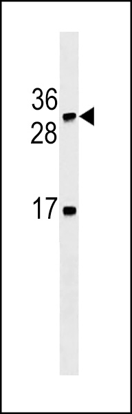
| Calculated MW | 25390 Da |
| Antigen Region | 199-227 aa |
| Gene ID | 4050 |
|---|---|
| Other Names | Lymphotoxin-beta, LT-beta, Tumor necrosis factor C, TNF-C, Tumor necrosis factor ligand superfamily member 3, LTB, TNFC, TNFSF3 |
| Target/Specificity | This LTB antibody is generated from mice immunized with a KLH conjugated synthetic peptide between 199-227 amino acids from the C-terminal region of human LTB. |
| Dilution | WB~~1:500~1000 E~~Use at an assay dependent concentration. |
| Format | Purified monoclonal antibody supplied in PBS with 0.09% (W/V) sodium azide. This antibody is prepared by Euglobin precipitation followed by dialysis against PBS. |
| Storage | Maintain refrigerated at 2-8°C for up to 2 weeks. For long term storage store at -20°C in small aliquots to prevent freeze-thaw cycles. |
| Precautions | LTB Antibody (C-term) is for research use only and not for use in diagnostic or therapeutic procedures. |
| Name | LTB |
|---|---|
| Synonyms | TNFC, TNFSF3 |
| Function | Cytokine that binds to LTBR/TNFRSF3 (PubMed:24248355). May play a specific role in immune response regulation. Provides the membrane anchor for the attachment of the heterotrimeric complex to the cell surface. Isoform 2 is probably non-functional. |
| Cellular Location | Membrane; Single-pass type II membrane protein |
| Tissue Location | Spleen and thymus. |

Thousands of laboratories across the world have published research that depended on the performance of antibodies from Abcepta to advance their research. Check out links to articles that cite our products in major peer-reviewed journals, organized by research category.
info@abcepta.com, and receive a free "I Love Antibodies" mug.

Provided below are standard protocols that you may find useful for product applications.
Background
Lymphotoxin beta is a type II membrane protein of the TNF family. It anchors lymphotoxin-alpha to the cell surface through heterotrimer formation. The predominant form on the lymphocyte surface is the lymphotoxin-alpha 1/beta 2 complex (e.g. 1 molecule alpha/2 molecules beta) and this complex is the primary ligand for the lymphotoxin-beta receptor. The minor complex is lymphotoxin-alpha 2/beta 1. LTB is an inducer of the inflammatory response system and involved in normal development of lymphoid tissue. Lymphotoxin-beta isoform b is unable to complex with lymphotoxin-alpha suggesting a function for lymphotoxin-beta which is independent of lympyhotoxin-alpha. Alternative splicing results in multiple transcript variants encoding different isoforms.
References
Clancy, R.M., et al. Arthritis Rheum. 62(11):3415-3424(2010)
Bailey, S.D., et al. Diabetes Care 33(10):2250-2253(2010)
Young, J., et al. Cytokine 51(1):78-86(2010)
McGeachie, M., et al. Circulation 120(24):2448-2454(2009)
Talmud, P.J., et al. Am. J. Hum. Genet. 85(5):628-642(2009)
If you have used an Abcepta product and would like to share how it has performed, please click on the "Submit Review" button and provide the requested information. Our staff will examine and post your review and contact you if needed.
If you have any additional inquiries please email technical services at tech@abcepta.com.
Ordering Information
Other Products
Shipping Information
