


 Foundational characteristics of cancer include proliferation, angiogenesis, migration, evasion of apoptosis, and cellular immortality. Find key markers for these cellular processes and antibodies to detect them.
 The SUMOplot™ Analysis Program predicts and scores sumoylation sites in your protein. SUMOylation is a post-translational modification involved in various cellular processes, such as nuclear-cytosolic transport, transcriptional regulation, apoptosis, protein stability, response to stress, and progression through the cell cycle.
 The Autophagy Receptor Motif Plotter predicts and scores autophagy receptor binding sites in your protein. Identifying proteins connected to this pathway is critical to understanding the role of autophagy in physiological as well as pathological processes such as development, differentiation, neurodegenerative diseases, stress, infection, and cancer.
NFKBIA Antibody
Mouse Monoclonal Antibody (Mab)
- SPECIFICATION
- CITATIONS
- PROTOCOLS
- BACKGROUND

Application
| WB, E |
|---|---|
| Primary Accession | P25963 |
| Reactivity | Human |
| Host | Mouse |
| Clonality | Monoclonal |
| Isotype | IgG1,κ |
| Clone/Animal Names | 1121CT8.6.1 |
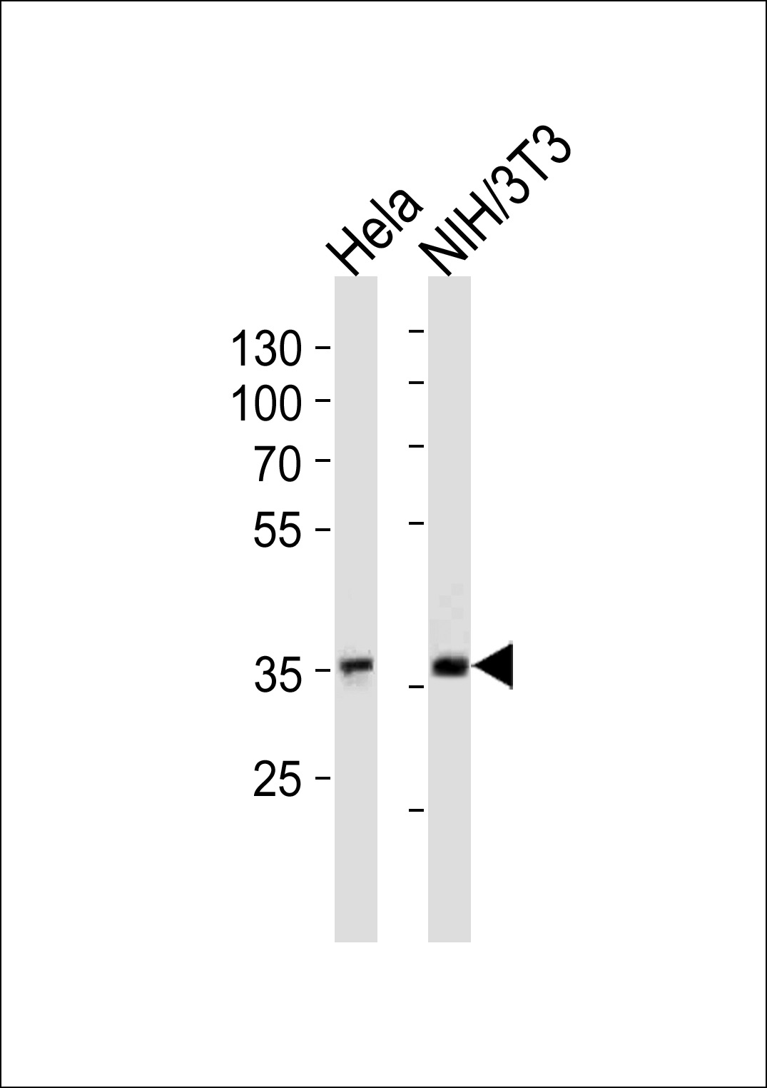
| Calculated MW | 35609 Da |
| Gene ID | 4792 |
|---|---|
| Other Names | NF-kappa-B inhibitor alpha, I-kappa-B-alpha, IkB-alpha, IkappaBalpha, Major histocompatibility complex enhancer-binding protein MAD3, NFKBIA, IKBA, MAD3, NFKBI |
| Target/Specificity | Purified His-tagged NFKBIA protein was used to produced this monoclonal antibody. |
| Dilution | WB~~1:1000 E~~Use at an assay dependent concentration. |
| Format | Purified monoclonal antibody supplied in PBS with 0.09% (W/V) sodium azide. This antibody is purified through a protein G column, followed by dialysis against PBS. |
| Storage | Maintain refrigerated at 2-8°C for up to 2 weeks. For long term storage store at -20°C in small aliquots to prevent freeze-thaw cycles. |
| Precautions | NFKBIA Antibody is for research use only and not for use in diagnostic or therapeutic procedures. |
| Name | NFKBIA |
|---|---|
| Synonyms | IKBA, MAD3, NFKBI |
| Function | Inhibits the activity of dimeric NF-kappa-B/REL complexes by trapping REL (RELA/p65 and NFKB1/p50) dimers in the cytoplasm by masking their nuclear localization signals (PubMed:1493333, PubMed:36651806, PubMed:7479976). On cellular stimulation by immune and pro-inflammatory responses, becomes phosphorylated promoting ubiquitination and degradation, enabling the dimeric RELA to translocate to the nucleus and activate transcription (PubMed:7479976, PubMed:7628694, PubMed:7796813, PubMed:7878466). |
| Cellular Location | Cytoplasm. Nucleus. Note=Shuttles between the nucleus and the cytoplasm by a nuclear localization signal (NLS) and a CRM1-dependent nuclear export. |

Thousands of laboratories across the world have published research that depended on the performance of antibodies from Abcepta to advance their research. Check out links to articles that cite our products in major peer-reviewed journals, organized by research category.
info@abcepta.com, and receive a free "I Love Antibodies" mug.

Provided below are standard protocols that you may find useful for product applications.
Background
Inhibits the activity of dimeric NF-kappa-B/REL complexes by trapping REL dimers in the cytoplasm through masking of their nuclear localization signals. On cellular stimulation by immune and proinflammatory responses, becomes phosphorylated promoting ubiquitination and degradation, enabling the dimeric RELA to translocate to the nucleus and activate transcription.
References
Huxford T., et al. Cell 95:759-770(1998).
Cockman M.E., et al. Proc. Natl. Acad. Sci. U.S.A. 103:14767-14772(2006).
Haskill S., et al. Cell 65:1281-1289(1991).
Jungnickel B., et al. J. Exp. Med. 191:395-402(2000).
Liu B., et al. Submitted (APR-2001) to the EMBL/GenBank/DDBJ databases.
If you have used an Abcepta product and would like to share how it has performed, please click on the "Submit Review" button and provide the requested information. Our staff will examine and post your review and contact you if needed.
If you have any additional inquiries please email technical services at tech@abcepta.com.
Ordering Information
Other Products
Shipping Information
