


 Foundational characteristics of cancer include proliferation, angiogenesis, migration, evasion of apoptosis, and cellular immortality. Find key markers for these cellular processes and antibodies to detect them.
 The SUMOplot™ Analysis Program predicts and scores sumoylation sites in your protein. SUMOylation is a post-translational modification involved in various cellular processes, such as nuclear-cytosolic transport, transcriptional regulation, apoptosis, protein stability, response to stress, and progression through the cell cycle.
 The Autophagy Receptor Motif Plotter predicts and scores autophagy receptor binding sites in your protein. Identifying proteins connected to this pathway is critical to understanding the role of autophagy in physiological as well as pathological processes such as development, differentiation, neurodegenerative diseases, stress, infection, and cancer.
BCL2L1 Antibody
Mouse Monoclonal Antibody (Mab)
- SPECIFICATION
- CITATIONS
- PROTOCOLS
- BACKGROUND

Application
| WB, E |
|---|---|
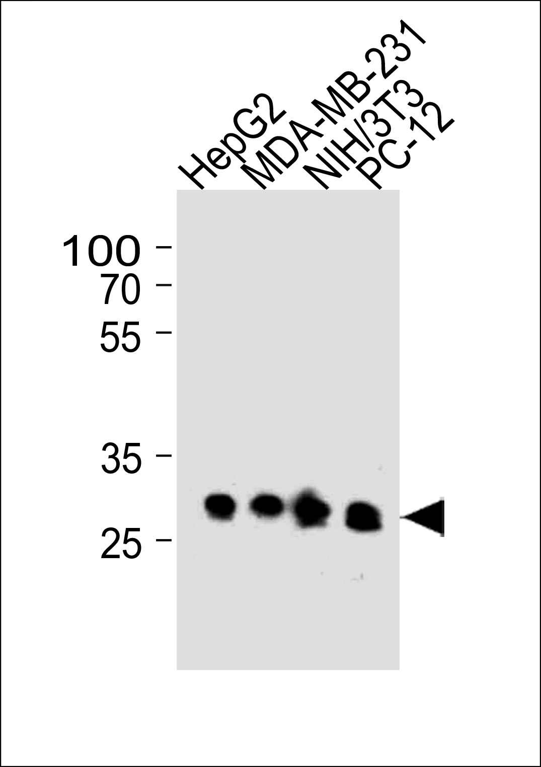
| Primary Accession | Q07817 |
| Reactivity | Human, Mouse, Rat |
| Host | Mouse |
| Clonality | Monoclonal |
| Isotype | IgG1 |
| Clone/Animal Names | 804CT19.1.4 |
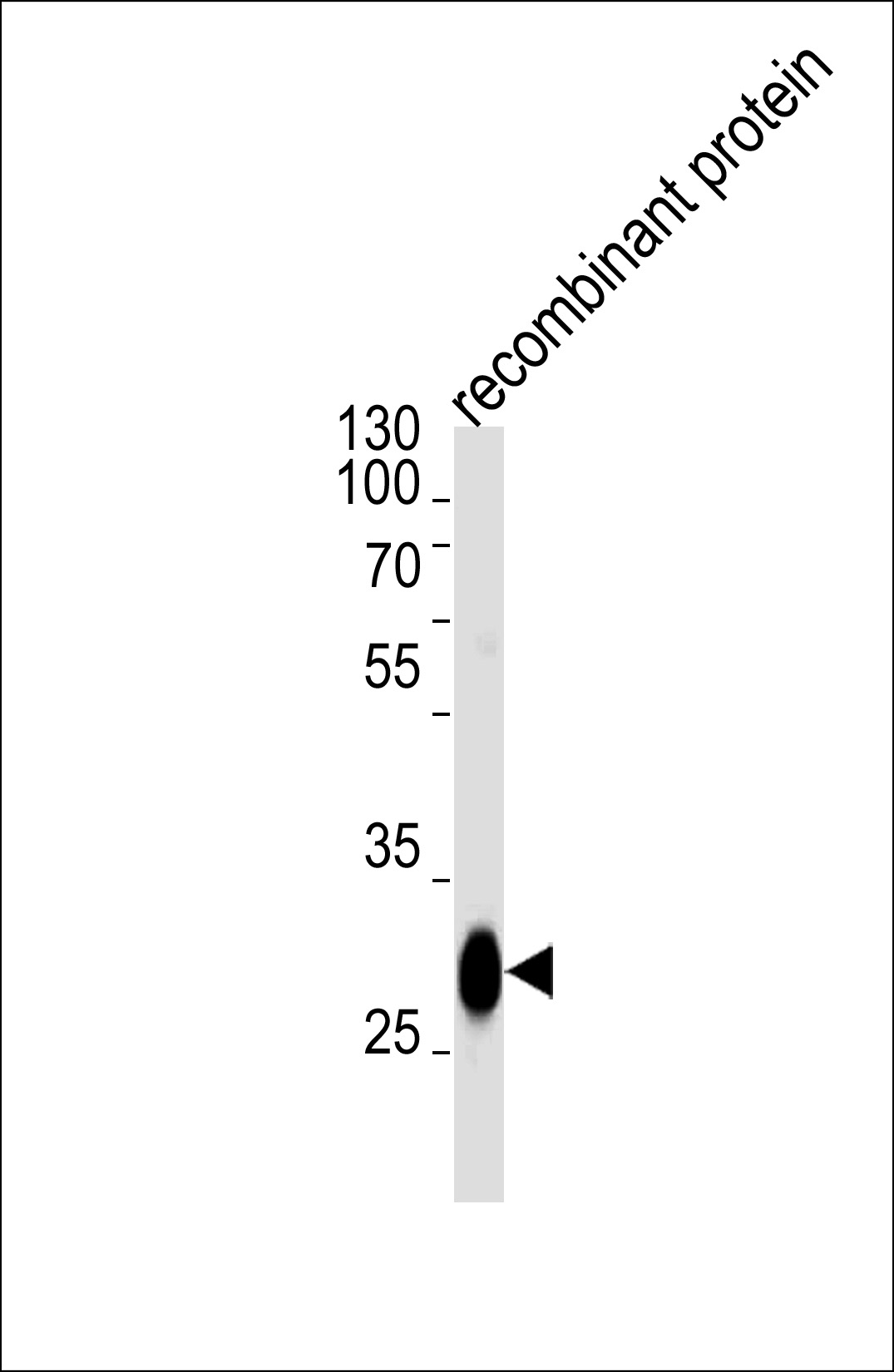
| Calculated MW | 26049 Da |
| Gene ID | 598 |
|---|---|
| Other Names | Bcl-2-like protein 1, Bcl2-L-1, Apoptosis regulator Bcl-X, BCL2L1, BCL2L, BCLX |
| Target/Specificity | Purified His-tagged BCL2L1 protein was used to produced this monoclonal antibody. |
| Dilution | WB~~1:1000 E~~Use at an assay dependent concentration. |
| Format | Purified monoclonal antibody supplied in PBS with 0.09% (W/V) sodium azide. This antibody is purified through a protein G column, followed by dialysis against PBS. |
| Storage | Maintain refrigerated at 2-8°C for up to 2 weeks. For long term storage store at -20°C in small aliquots to prevent freeze-thaw cycles. |
| Precautions | BCL2L1 Antibody is for research use only and not for use in diagnostic or therapeutic procedures. |
| Name | BCL2L1 |
|---|---|
| Synonyms | BCL2L, BCLX |
| Function | Potent inhibitor of cell death. Inhibits activation of caspases. Appears to regulate cell death by blocking the voltage- dependent anion channel (VDAC) by binding to it and preventing the release of the caspase activator, CYC1, from the mitochondrial membrane. Also acts as a regulator of G2 checkpoint and progression to cytokinesis during mitosis. Isoform Bcl-X(S) promotes apoptosis. |
| Cellular Location | [Isoform Bcl-X(L)]: Mitochondrion inner membrane. Mitochondrion outer membrane Mitochondrion matrix. Cytoplasmic vesicle, secretory vesicle, synaptic vesicle membrane. Cytoplasm, cytosol. Cytoplasm, cytoskeleton, microtubule organizing center, centrosome. Nucleus membrane; Single-pass membrane protein; Cytoplasmic side. Note=After neuronal stimulation, translocates from cytosol to synaptic vesicle and mitochondrion membrane in a calmodulin-dependent manner (By similarity). Localizes to the centrosome when phosphorylated at Ser-49 |
| Tissue Location | Bcl-X(S) is expressed at high levels in cells that undergo a high rate of turnover, such as developing lymphocytes. In contrast, Bcl-X(L) is found in tissues containing long-lived postmitotic cells, such as adult brain |

Thousands of laboratories across the world have published research that depended on the performance of antibodies from Abcepta to advance their research. Check out links to articles that cite our products in major peer-reviewed journals, organized by research category.
info@abcepta.com, and receive a free "I Love Antibodies" mug.

Provided below are standard protocols that you may find useful for product applications.
Background
Potent inhibitor of cell death. Inhibits activation of caspases (By similarity). Appears to regulate cell death by blocking the voltage-dependent anion channnel (VDAC) by binding to it and preventing the release of the caspase activator, CYC1, from the mitochondrial membrane. Also acts as a regulator of G2 checkpoint and progression to cytokinesis during mitosis. Isoform Bcl-X(S) promotes apoptosis.
References
Boise L.H., et al. Cell 74:597-608(1993).
Ban J., et al. Biochem. Biophys. Res. Commun. 248:147-152(1998).
Inohara N., et al. Submitted (OCT-1996) to the EMBL/GenBank/DDBJ databases.
Bechtel S., et al. BMC Genomics 8:399-399(2007).
Kalnine N., et al. Submitted (OCT-2004) to the EMBL/GenBank/DDBJ databases.
If you have used an Abcepta product and would like to share how it has performed, please click on the "Submit Review" button and provide the requested information. Our staff will examine and post your review and contact you if needed.
If you have any additional inquiries please email technical services at tech@abcepta.com.
Ordering Information
Other Products
Shipping Information
