


 Foundational characteristics of cancer include proliferation, angiogenesis, migration, evasion of apoptosis, and cellular immortality. Find key markers for these cellular processes and antibodies to detect them.
 The SUMOplot™ Analysis Program predicts and scores sumoylation sites in your protein. SUMOylation is a post-translational modification involved in various cellular processes, such as nuclear-cytosolic transport, transcriptional regulation, apoptosis, protein stability, response to stress, and progression through the cell cycle.
 The Autophagy Receptor Motif Plotter predicts and scores autophagy receptor binding sites in your protein. Identifying proteins connected to this pathway is critical to understanding the role of autophagy in physiological as well as pathological processes such as development, differentiation, neurodegenerative diseases, stress, infection, and cancer.
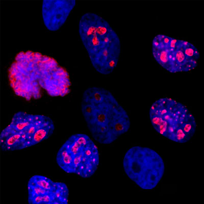
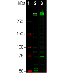
Anti-Ki-67 Antibody
Our Anti-Ki-67 Antibody primary antibody from PhosphoSolutions is mouse monoclonal. It detects human
- SPECIFICATION
- CITATIONS
- PROTOCOLS
- BACKGROUND

Application
| WB, IHC |
|---|---|
| Primary Accession | P46013 |
| Host | Mouse |
| Clonality | Monoclonal |
| Isotype | IgG1 |
| Clone Names | 6G3 |
| Calculated MW | 358694 Da |
| Gene ID | 4288 |
|---|---|
| Other Names | Antigen identified by monoclonal antibody Ki 67 antibody, Antigen identified by monoclonal antibody Ki-67 antibody, Antigen KI-67 antibody, Antigen KI67 antibody, Antigen Ki67 antibody, Ki67 antibody, KI67_HUMAN antibody, KIA antibody, Marker of proliferation Ki-67 antibody, MIB 1 antibody, MIB antibody, MKI67 antibody, PPP1R105 antibody, Proliferation marker protein Ki-67 antibody, Proliferation related Ki 67 antigen antibody, Protein phosphatase 1 regulatory subunit 105 antibody, RP11-380J17.2 antibody |
| Target/Specificity | Ki-67 is a nuclear protein that is considered to be essential for cellular proliferation since it is expressed only in active phases of the cell cycle (G1, S, G2 and mitosis) but not in resting phase G0 (Scholzen & Gerdes 2000). Ki-67 antibodies have therefore been widely used as markers of cell growth. Ki-67 has further been identified as a predicative and prognostic marker in breast, lung, cervical and other cancers (Yerushalmi et al., 2010; Folescu et al., 2018; Das et al., 2018). |
| Dilution | WB~~1:1000 IHC~~1:100~500 |
| Format | Protein G Purified |
| Storage | Maintain refrigerated at 2-8°C for up to 6 months. For long term storage store at -20°C in small aliquots to prevent freeze-thaw cycles. |
| Precautions | Anti-Ki-67 Antibody is for research use only and not for use in diagnostic or therapeutic procedures. |
| Shipping | Blue Ice |

Thousands of laboratories across the world have published research that depended on the performance of antibodies from Abcepta to advance their research. Check out links to articles that cite our products in major peer-reviewed journals, organized by research category.
info@abcepta.com, and receive a free "I Love Antibodies" mug.

Provided below are standard protocols that you may find useful for product applications.
Background
Ki-67 is a nuclear protein that is considered to be essential for cellular proliferation since it is expressed only in active phases of the cell cycle (G1, S, G2 and mitosis) but not in resting phase G0 (Scholzen & Gerdes 2000). Ki-67 antibodies have therefore been widely used as markers of cell growth. Ki-67 has further been identified as a predicative and prognostic marker in breast, lung, cervical and other cancers (Yerushalmi et al., 2010; Folescu et al., 2018; Das et al., 2018).
If you have used an Abcepta product and would like to share how it has performed, please click on the "Submit Review" button and provide the requested information. Our staff will examine and post your review and contact you if needed.
If you have any additional inquiries please email technical services at tech@abcepta.com.
Ordering Information
Other Products
Shipping Information
