


 Foundational characteristics of cancer include proliferation, angiogenesis, migration, evasion of apoptosis, and cellular immortality. Find key markers for these cellular processes and antibodies to detect them.
 The SUMOplot™ Analysis Program predicts and scores sumoylation sites in your protein. SUMOylation is a post-translational modification involved in various cellular processes, such as nuclear-cytosolic transport, transcriptional regulation, apoptosis, protein stability, response to stress, and progression through the cell cycle.
 The Autophagy Receptor Motif Plotter predicts and scores autophagy receptor binding sites in your protein. Identifying proteins connected to this pathway is critical to understanding the role of autophagy in physiological as well as pathological processes such as development, differentiation, neurodegenerative diseases, stress, infection, and cancer.
Anti-NSE (Neuron specific enolase) Antibody
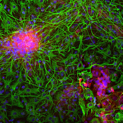
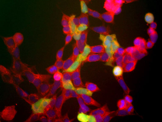
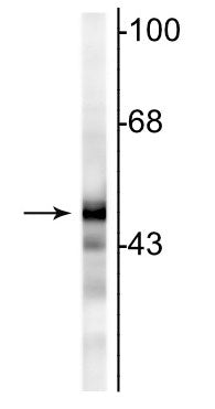
Our Anti-NSE (Neuron specific enolase) primary antibody from PhosphoSolutions is rabbit polyclonal.
- SPECIFICATION
- CITATIONS
- PROTOCOLS
- BACKGROUND

Application
| WB, IHC |
|---|---|
| Primary Accession | P09104 |
| Reactivity | Bovine, Chicken, Drosophila |
| Host | Rabbit |
| Clonality | Polyclonal |
| Isotype | IgG |
| Calculated MW | 47269 Da |
| Gene ID | 2026 |
|---|---|
| Other Names | 2 phospho D glycerate hydrolyase antibody, 2-phospho-D-glycerate hydro-lyase antibody, Eno 2 antibody, ENO2 antibody, ENOG antibody, ENOG_HUMAN antibody, Enolase 2 (gamma, neuronal) antibody, Enolase 2 antibody, Enolase 2 gamma neuronal antibody, Enolase2 antibody, Epididymis secretory protein Li 279 antibody, Gamma enolase antibody, Gamma-enolase antibody, HEL S 279 antibody, Neural enolase antibody, Neuron specific enolase antibody, Neuron specific gamma enolase antibody, Neuron-specific enolase antibody, neuronal enriched enolase antibody, Neurone specific enolase antibody, NSE antibody |
| Target/Specificity | Neuron specific enolase (NSE) is an enzyme which catalyzes the conversion of 2-phosphoglycerate to phosphoenolpyruvate in the glycolytic pathway, and also the reverse reaction in gluconeogenesis. It is one of three mammalian enolases, which are also known as ENO1, ENO2, and ENO3 or alternately as enolase alpha, beta and gamma. The three enolases have different cell type specific expression patterns, so that antibodies to them are useful cell type specific markers.(MacAlesse et al., 1988). NSE corresponds to ENO2 or enolase gamma and is heavily expressed in neuronal cells. ENO1 is also known as enolase alpha and as non-neuronal enolase. The third enolase, ENO3 or enolase beta, is expressed in muscle cells. Since neurons require a great deal of energy, they are very rich in glycolytic enzymes such a GAPDH and NSE. Antibodies to this protein are therefore useful to identify neuronal cell bodies, developing neuronal lineage and neuroendocrine cells. Release of NSE from damaged neurons into CSF and blood has also been used as a biomarker of neuronal injury (2). |
| Dilution | WB~~1:1000 IHC~~1:100~500 |
| Format | Neat Serum |
| Storage | Maintain refrigerated at 2-8°C for up to 6 months. For long term storage store at -20°C in small aliquots to prevent freeze-thaw cycles. |
| Precautions | Anti-NSE (Neuron specific enolase) Antibody is for research use only and not for use in diagnostic or therapeutic procedures. |
| Shipping | Blue Ice |

Thousands of laboratories across the world have published research that depended on the performance of antibodies from Abcepta to advance their research. Check out links to articles that cite our products in major peer-reviewed journals, organized by research category.
info@abcepta.com, and receive a free "I Love Antibodies" mug.

Provided below are standard protocols that you may find useful for product applications.
Background
Neuron specific enolase (NSE) is an enzyme which catalyzes the conversion of 2-phosphoglycerate to phosphoenolpyruvate in the glycolytic pathway, and also the reverse reaction in gluconeogenesis. It is one of three mammalian enolases, which are also known as ENO1, ENO2, and ENO3 or alternately as enolase alpha, beta and gamma. The three enolases have different cell type specific expression patterns, so that antibodies to them are useful cell type specific markers.(MacAlesse et al., 1988). NSE corresponds to ENO2 or enolase gamma and is heavily expressed in neuronal cells. ENO1 is also known as enolase alpha and as non-neuronal enolase. The third enolase, ENO3 or enolase beta, is expressed in muscle cells. Since neurons require a great deal of energy, they are very rich in glycolytic enzymes such a GAPDH and NSE. Antibodies to this protein are therefore useful to identify neuronal cell bodies, developing neuronal lineage and neuroendocrine cells. Release of NSE from damaged neurons into CSF and blood has also been used as a biomarker of neuronal injury (2).
If you have used an Abcepta product and would like to share how it has performed, please click on the "Submit Review" button and provide the requested information. Our staff will examine and post your review and contact you if needed.
If you have any additional inquiries please email technical services at tech@abcepta.com.
Ordering Information
Other Products
Shipping Information
