


 Foundational characteristics of cancer include proliferation, angiogenesis, migration, evasion of apoptosis, and cellular immortality. Find key markers for these cellular processes and antibodies to detect them.
 The SUMOplot™ Analysis Program predicts and scores sumoylation sites in your protein. SUMOylation is a post-translational modification involved in various cellular processes, such as nuclear-cytosolic transport, transcriptional regulation, apoptosis, protein stability, response to stress, and progression through the cell cycle.
 The Autophagy Receptor Motif Plotter predicts and scores autophagy receptor binding sites in your protein. Identifying proteins connected to this pathway is critical to understanding the role of autophagy in physiological as well as pathological processes such as development, differentiation, neurodegenerative diseases, stress, infection, and cancer.
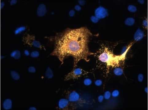
Anti-SARM1 Antibody
Our Anti-SARM1 Antibody primary antibody from PhosphoSolutions is rabbit polyclonal. It detects huma
- SPECIFICATION
- CITATIONS
- PROTOCOLS
- BACKGROUND

Application
| WB |
|---|---|
| Primary Accession | Q6SZW1 |
| Reactivity | Bovine |
| Host | Rabbit |
| Clonality | Polyclonal |
| Isotype | IgG |
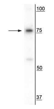
| Calculated MW | 79388 Da |
| Gene ID | 23098 |
|---|---|
| Other Names | FLJ36296 antibody, KIAA0524 antibody, MyD88-5 antibody, SAM and ARM-containing protein antibody, SAM domain-containing protein 2 antibody, SAMD2 antibody, SARM 1 antibody, SARM1 antibody, SARM1_HUMAN antibody, Sterile alpha and Armadillo repeat protein antibody, sterile alpha and HEAT/Armadillo motif protein, ortholog of Drosophila antibody, sterile alpha and HEAT/Armadillo motifs-containing protein antibody, sterile alpha and TIR motif containing 1 antibody, Sterile alpha and TIR motif-containing protein 1 antibody, Sterile alpha and TIR motifs-containing protein 1 antibody, Sterile alpha motif domain-containing protein 2 antibody, Tir-1 homolog antibody |
| Target/Specificity | NAD(+) hydrolase sterile alpha and TIR motif-containing protein 1 (SARM1), is the fifth member of the Toll/Il-1 Receptor (TIR) domain protein family. The highly conserved 724 amino acid protein is predominantly expressed in the brain, kidney and liver (Mink, M., et al 2001). Furthermore, SARM1 is enriched in the cytoplasm, mitchondria, and the axons, dendrites, and synapses of neurons (Panneerselvam, P., et al, 2012). SARM1 plays a key role in axonal degeneration by catalyzing the cleavage of NAD+ into ADPR, cADPR, and nicotinamide in response to injury triggering Wallerian degeneration (Gerdts, J. et al 2015). SARM1 has also been shown to activate neuronal cell death in response to stress (Peng, J. et al. 2010). |
| Dilution | WB~~1:1000 |
| Format | Antigen Affinity Purified from Pooled Serum |
| Storage | Maintain refrigerated at 2-8°C for up to 6 months. For long term storage store at -20°C in small aliquots to prevent freeze-thaw cycles. |
| Precautions | Anti-SARM1 Antibody is for research use only and not for use in diagnostic or therapeutic procedures. |
| Shipping | Blue Ice |

Thousands of laboratories across the world have published research that depended on the performance of antibodies from Abcepta to advance their research. Check out links to articles that cite our products in major peer-reviewed journals, organized by research category.
info@abcepta.com, and receive a free "I Love Antibodies" mug.

Provided below are standard protocols that you may find useful for product applications.
Background
NAD(+) hydrolase sterile alpha and TIR motif-containing protein 1 (SARM1), is the fifth member of the Toll/Il-1 Receptor (TIR) domain protein family. The highly conserved 724 amino acid protein is predominantly expressed in the brain, kidney and liver (Mink, M., et al 2001). Furthermore, SARM1 is enriched in the cytoplasm, mitchondria, and the axons, dendrites, and synapses of neurons (Panneerselvam, P., et al, 2012). SARM1 plays a key role in axonal degeneration by catalyzing the cleavage of NAD+ into ADPR, cADPR, and nicotinamide in response to injury triggering Wallerian degeneration (Gerdts, J. et al 2015). SARM1 has also been shown to activate neuronal cell death in response to stress (Peng, J. et al. 2010).
If you have used an Abcepta product and would like to share how it has performed, please click on the "Submit Review" button and provide the requested information. Our staff will examine and post your review and contact you if needed.
If you have any additional inquiries please email technical services at tech@abcepta.com.
Ordering Information
Other Products
Shipping Information
