


 Foundational characteristics of cancer include proliferation, angiogenesis, migration, evasion of apoptosis, and cellular immortality. Find key markers for these cellular processes and antibodies to detect them.
 The SUMOplot™ Analysis Program predicts and scores sumoylation sites in your protein. SUMOylation is a post-translational modification involved in various cellular processes, such as nuclear-cytosolic transport, transcriptional regulation, apoptosis, protein stability, response to stress, and progression through the cell cycle.
 The Autophagy Receptor Motif Plotter predicts and scores autophagy receptor binding sites in your protein. Identifying proteins connected to this pathway is critical to understanding the role of autophagy in physiological as well as pathological processes such as development, differentiation, neurodegenerative diseases, stress, infection, and cancer.
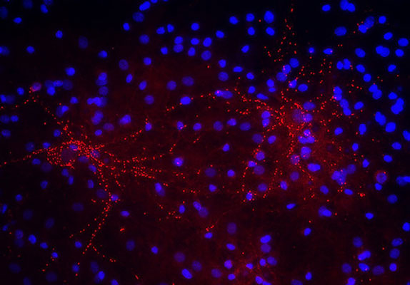
Anti-Synaptophysin 2 Antibody
Our Anti-Synaptophysin 2 rabbit polyclonal primary antibody from PhosphoSolutions is produced in-hou
- SPECIFICATION
- CITATIONS
- PROTOCOLS
- BACKGROUND

Application
| WB |
|---|---|
| Primary Accession | P22831 |
| Host | Rabbit |
| Clonality | Polyclonal |
| Isotype | IgG |
| Calculated MW | 29107 Da |
| Gene ID | 66030 |
|---|---|
| Other Names | DKFZp686G0883 antibody, MGC26651 antibody, SPO antibody, Synaptophysin 2 antibody, Synaptoporin antibody, Synpr antibody, SYNPR_HUMAN antibody |
| Target/Specificity | Synaptophysin 2, also known as Synaptoporin, is a component of the synaptic vesicle membrane and thought to play an important role in synaptic vesicle trafficking (Dai et al., 2003). It is highly homologous to Synaptophysin 1, yet differs in expression. Whereas Synaptophysin 1 is ubiquitously expressed, Synaptophysin 2 is concentrated in the mossy fiber synapses of the hippocampus (Grabs et al., 1994; Singec et al., 2002). |
| Dilution | WB~~1:1000 |
| Format | Antigen Affinity Purified from Pooled Serum |
| Storage | Maintain refrigerated at 2-8°C for up to 6 months. For long term storage store at -20°C in small aliquots to prevent freeze-thaw cycles. |
| Precautions | Anti-Synaptophysin 2 Antibody is for research use only and not for use in diagnostic or therapeutic procedures. |
| Shipping | Blue Ice |

Thousands of laboratories across the world have published research that depended on the performance of antibodies from Abcepta to advance their research. Check out links to articles that cite our products in major peer-reviewed journals, organized by research category.
info@abcepta.com, and receive a free "I Love Antibodies" mug.

Provided below are standard protocols that you may find useful for product applications.
Background
Synaptophysin 2, also known as Synaptoporin, is a component of the synaptic vesicle membrane and thought to play an important role in synaptic vesicle trafficking (Dai et al., 2003). It is highly homologous to Synaptophysin 1, yet differs in expression. Whereas Synaptophysin 1 is ubiquitously expressed, Synaptophysin 2 is concentrated in the mossy fiber synapses of the hippocampus (Grabs et al., 1994; Singec et al., 2002).
If you have used an Abcepta product and would like to share how it has performed, please click on the "Submit Review" button and provide the requested information. Our staff will examine and post your review and contact you if needed.
If you have any additional inquiries please email technical services at tech@abcepta.com.
Ordering Information
Shipping Information
