


 Foundational characteristics of cancer include proliferation, angiogenesis, migration, evasion of apoptosis, and cellular immortality. Find key markers for these cellular processes and antibodies to detect them.
 The SUMOplot™ Analysis Program predicts and scores sumoylation sites in your protein. SUMOylation is a post-translational modification involved in various cellular processes, such as nuclear-cytosolic transport, transcriptional regulation, apoptosis, protein stability, response to stress, and progression through the cell cycle.
 The Autophagy Receptor Motif Plotter predicts and scores autophagy receptor binding sites in your protein. Identifying proteins connected to this pathway is critical to understanding the role of autophagy in physiological as well as pathological processes such as development, differentiation, neurodegenerative diseases, stress, infection, and cancer.
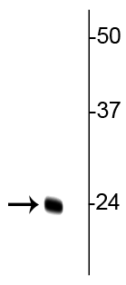
Anti-TFAM (Transcription Factor A, mitochondrial) Antibody
Our Anti-TFAM (Transcription Factor A, mitochondrial) primary antibody from PhosphoSolutions is rabb
- SPECIFICATION
- CITATIONS
- PROTOCOLS
- BACKGROUND

| Primary Accession | P40630 |
|---|---|
| Reactivity | Bovine |
| Host | Rabbit |
| Clonality | Polyclonal |
| Isotype | IgG |
| Calculated MW | 27988 Da |
| Gene ID | 21780 |
|---|---|
| Other Names | Transcription factor 6-like 1 antibody, Mitochondrial transcription factor 1 antibody, mitochondrial transcription factor A antibody, MtTF1 antibody, mtTFA antibody, TCF 6 antibody, TCF-6 antibody, TCF6 antibody, TCF6L1 antibody, TCF6L2 antibody, TCF6L3 antibody, TFAM antibody, TFAM_HUMAN antibody, Transcription factor 6 antibody, Transcription factor 6 like 2 (mitochondrial transcription factor) antibody, Transcription factor 6 like 2 antibody, Transcription factor 6-like 2 antibody, transcription factor 6-like 3 antibody, Transcription factor A mitochondrial antibody, Transcription factor A mitochondrial antibody, Transcription factor A mitochondrial precursor antibody |
| Target/Specificity | Mitochondrial Transcription Factor A (TFAM) is a key activator of mitochondrial (mt) DNA transcription as well as a participant in mitochondrial genome replication. mtDNA is highly susceptible to oxidative stress leading to mitochondrial dysfunction. Overexpression of TFAM has been implicated in the amelioration of age dependent impairment of brain functions through the prevention of oxidative stress and mitochondrial dysfunction in microglia (Hayashi et al., 2008). More recently, TFAM overexpression has been shown to potentially reduce oxidative stress in motor neurons and delay onset of amyotrophic lateral sclerosis (ALS) in ALS model mice (Morimoto et al., 2012). |
| Format | Neat Serum |
| Storage | Maintain refrigerated at 2-8°C for up to 6 months. For long term storage store at -20°C in small aliquots to prevent freeze-thaw cycles. |
| Precautions | Anti-TFAM (Transcription Factor A, mitochondrial) Antibody is for research use only and not for use in diagnostic or therapeutic procedures. |
| Shipping | Blue Ice |

Thousands of laboratories across the world have published research that depended on the performance of antibodies from Abcepta to advance their research. Check out links to articles that cite our products in major peer-reviewed journals, organized by research category.
info@abcepta.com, and receive a free "I Love Antibodies" mug.

Provided below are standard protocols that you may find useful for product applications.
Background
Mitochondrial Transcription Factor A (TFAM) is a key activator of mitochondrial (mt) DNA transcription as well as a participant in mitochondrial genome replication. mtDNA is highly susceptible to oxidative stress leading to mitochondrial dysfunction. Overexpression of TFAM has been implicated in the amelioration of age dependent impairment of brain functions through the prevention of oxidative stress and mitochondrial dysfunction in microglia (Hayashi et al., 2008). More recently, TFAM overexpression has been shown to potentially reduce oxidative stress in motor neurons and delay onset of amyotrophic lateral sclerosis (ALS) in ALS model mice (Morimoto et al., 2012).
If you have used an Abcepta product and would like to share how it has performed, please click on the "Submit Review" button and provide the requested information. Our staff will examine and post your review and contact you if needed.
If you have any additional inquiries please email technical services at tech@abcepta.com.
Ordering Information
Other Products
Shipping Information
