


 Foundational characteristics of cancer include proliferation, angiogenesis, migration, evasion of apoptosis, and cellular immortality. Find key markers for these cellular processes and antibodies to detect them.
 The SUMOplot™ Analysis Program predicts and scores sumoylation sites in your protein. SUMOylation is a post-translational modification involved in various cellular processes, such as nuclear-cytosolic transport, transcriptional regulation, apoptosis, protein stability, response to stress, and progression through the cell cycle.
 The Autophagy Receptor Motif Plotter predicts and scores autophagy receptor binding sites in your protein. Identifying proteins connected to this pathway is critical to understanding the role of autophagy in physiological as well as pathological processes such as development, differentiation, neurodegenerative diseases, stress, infection, and cancer.
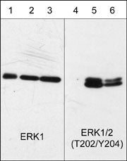
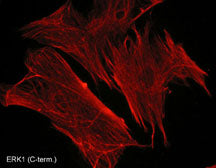
Anti-ERK1 (C-terminal region) Antibody
- SPECIFICATION
- CITATIONS
- PROTOCOLS
- BACKGROUND

Application
| WB, IHC |
|---|---|
| Primary Accession | P28482 |
| Reactivity | Bovine |
| Host | Mouse |
| Clonality | Mouse Monoclonal |
| Isotype | IgG1 |
| Clone Names | M233 |
| Calculated MW | 41390 Da |
| Gene ID | 5594 |
|---|---|
| Other Names | ERK, p42, p44, MAPK |
| Target/Specificity | Mitogen-activated protein kinases (MAPKs) are a widely conserved family of serine/threonine protein kinases involved in many cellular programs such as cell proliferation, differentiation, motility, and death. The ERK1/2 (p44/42) signaling pathway can be activated in response to a diverse range of extracellular stimuli including mitogens, growth factors, and cytokines. Upon stimulation, a sequential three-part protein kinase cascade is initiated, consisting of a MAP kinase kinase kinase (MAPKKK), a MAP kinase kinase (MAPKK), and a MAP kinase (MAPK). Multiple ERK1/2 MAPKKKs have been identified, including members of the Raf family as well as Mos and Tpl2/Cot. MEK1 and MEK2 are the primary MAPKKs in this pathway. MEK1 and MEK2 activate ERK1 and ERK2 through phosphorylation of activation loop residues Thr-202/Tyr-204 and Thr-185/Tyr-187, respectively. ERK1/2 are negatively regulated by a family of dual-specificity (Thr/Tyr) MAPK phosphatases. Several downstream targets of ERK1/2 have been identified, including p90RSK and the transcription factor Elk-1. |
| Dilution | WB~~1:1000 IHC~~1:100~500 |
| Storage | Maintain refrigerated at 2-8°C for up to 6 months. For long term storage store at -20°C in small aliquots to prevent freeze-thaw cycles. |
| Precautions | Anti-ERK1 (C-terminal region) Antibody is for research use only and not for use in diagnostic or therapeutic procedures. |
| Shipping | Blue Ice |

Thousands of laboratories across the world have published research that depended on the performance of antibodies from Abcepta to advance their research. Check out links to articles that cite our products in major peer-reviewed journals, organized by research category.
info@abcepta.com, and receive a free "I Love Antibodies" mug.

Provided below are standard protocols that you may find useful for product applications.
Background
Mitogen-activated protein kinases (MAPKs) are a widely conserved family of serine/threonine protein kinases involved in many cellular programs such as cell proliferation, differentiation, motility, and death. The ERK1/2 (p44/42) signaling pathway can be activated in response to a diverse range of extracellular stimuli including mitogens, growth factors, and cytokines. Upon stimulation, a sequential three-part protein kinase cascade is initiated, consisting of a MAP kinase kinase kinase (MAPKKK), a MAP kinase kinase (MAPKK), and a MAP kinase (MAPK). Multiple ERK1/2 MAPKKKs have been identified, including members of the Raf family as well as Mos and Tpl2/Cot. MEK1 and MEK2 are the primary MAPKKs in this pathway. MEK1 and MEK2 activate ERK1 and ERK2 through phosphorylation of activation loop residues Thr-202/Tyr-204 and Thr-185/Tyr-187, respectively. ERK1/2 are negatively regulated by a family of dual-specificity (Thr/Tyr) MAPK phosphatases. Several downstream targets of ERK1/2 have been identified, including p90RSK and the transcription factor Elk-1.
If you have used an Abcepta product and would like to share how it has performed, please click on the "Submit Review" button and provide the requested information. Our staff will examine and post your review and contact you if needed.
If you have any additional inquiries please email technical services at tech@abcepta.com.
Ordering Information
Other Products
Shipping Information
