


 Foundational characteristics of cancer include proliferation, angiogenesis, migration, evasion of apoptosis, and cellular immortality. Find key markers for these cellular processes and antibodies to detect them.
 The SUMOplot™ Analysis Program predicts and scores sumoylation sites in your protein. SUMOylation is a post-translational modification involved in various cellular processes, such as nuclear-cytosolic transport, transcriptional regulation, apoptosis, protein stability, response to stress, and progression through the cell cycle.
 The Autophagy Receptor Motif Plotter predicts and scores autophagy receptor binding sites in your protein. Identifying proteins connected to this pathway is critical to understanding the role of autophagy in physiological as well as pathological processes such as development, differentiation, neurodegenerative diseases, stress, infection, and cancer.
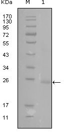
GATA3 Antibody
Purified Mouse Monoclonal Antibody
- SPECIFICATION
- CITATIONS
- PROTOCOLS
- BACKGROUND

Application
| WB, E |
|---|---|
| Primary Accession | P23771 |
| Reactivity | Human |
| Host | Mouse |
| Clonality | Monoclonal |
| Clone Names | 1A10D1 |
| Isotype | IgG1 |
| Calculated MW | 47916 Da |
| Description | GATA3: GATA binding protein 3. The genes for all 4 subunits of the T-cell antigen receptor (alpha, beta, gamma and delta) are controlled by distinct enhancers and their enhancer-binding proteins. Marine and Winoto (1991) identified a common TCR regulatory element by demonstrating binding of the enhancer-binding protein GATA3 to the enhancer elements of all 4 TCR genes. GATA3 had been shown in the chicken to be an enhancer-binding protein containing a zinc finger domain. GATA3 mRNA was demonstrated by Northern blot analysis in T cells but not in B cells, macrophages, or HeLa cell lines. GATA3 is abundantly expressed in the T-lymphocyte lineage and is thought to participate in T-cell receptor gene activation through binding to enhancers. Labastie et al. (1994) cloned the human gene and the 5-prime end of the mouse gene. The human gene comprises 6 exons distributed over 17 kb of DNA. Its 2 zinc fingers are encoded by 2 separate exons highly conserved with those of GATA1,but no other structural homologies between the 2 genes could be found. |
| Immunogen | Purified recombinant fragment of GATA3 (aa175-388) expressed in E. Coli. |
| Formulation | Ascitic fluid containing 0.03% sodium azide. |
| Gene ID | 2625 |
|---|---|
| Other Names | Trans-acting T-cell-specific transcription factor GATA-3, GATA-binding factor 3, GATA3 |
| Dilution | WB~~1/500 - 1/2000 E~~N/A |
| Storage | Maintain refrigerated at 2-8°C for up to 6 months. For long term storage store at -20°C in small aliquots to prevent freeze-thaw cycles. |
| Precautions | GATA3 Antibody is for research use only and not for use in diagnostic or therapeutic procedures. |
| Name | GATA3 |
|---|---|
| Function | Transcriptional activator which binds to the enhancer of the T-cell receptor alpha and delta genes. Binds to the consensus sequence 5'-AGATAG-3'. Required for the T-helper 2 (Th2) differentiation process following immune and inflammatory responses. Positively regulates ASB2 expression (By similarity). Coordinates macrophage transcriptional activation and UCP2-dependent metabolic reprogramming in response to IL33. Upon tissue injury, acts downstream of IL33 signaling to drive differentiation of inflammation-resolving alternatively activated macrophages. |
| Cellular Location | Nucleus. |
| Tissue Location | T-cells and endothelial cells. |

Thousands of laboratories across the world have published research that depended on the performance of antibodies from Abcepta to advance their research. Check out links to articles that cite our products in major peer-reviewed journals, organized by research category.
info@abcepta.com, and receive a free "I Love Antibodies" mug.

Provided below are standard protocols that you may find useful for product applications.
References
1. Cancer Res. 2005 Dec 15;65(24):11259-64. 2. J Histochem Cytochem. 2006 Feb;54(2):161-9. 3. Int Arch Allergy Immunol. 2006;139(4):306-16.
If you have used an Abcepta product and would like to share how it has performed, please click on the "Submit Review" button and provide the requested information. Our staff will examine and post your review and contact you if needed.
If you have any additional inquiries please email technical services at tech@abcepta.com.
Ordering Information
Other Products
Shipping Information
