


 Foundational characteristics of cancer include proliferation, angiogenesis, migration, evasion of apoptosis, and cellular immortality. Find key markers for these cellular processes and antibodies to detect them.
 The SUMOplot™ Analysis Program predicts and scores sumoylation sites in your protein. SUMOylation is a post-translational modification involved in various cellular processes, such as nuclear-cytosolic transport, transcriptional regulation, apoptosis, protein stability, response to stress, and progression through the cell cycle.
 The Autophagy Receptor Motif Plotter predicts and scores autophagy receptor binding sites in your protein. Identifying proteins connected to this pathway is critical to understanding the role of autophagy in physiological as well as pathological processes such as development, differentiation, neurodegenerative diseases, stress, infection, and cancer.
PAR4 Antibody
Purified Mouse Monoclonal Antibody
- SPECIFICATION
- CITATIONS
- PROTOCOLS
- BACKGROUND

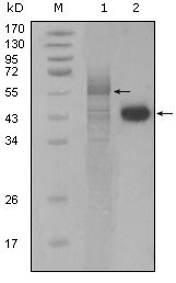
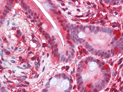
Application
| WB, IHC, E |
|---|---|
| Primary Accession | Q96IZ0 |
| Reactivity | Human |
| Host | Mouse |
| Clonality | Monoclonal |
| Clone Names | 3G9H7; 4H12E9 |
| Isotype | IgG1 |
| Calculated MW | 36568 Da |
| Description | Prostate apoptosis response 4 (Par4) is a 38kD protein originally identified as the product of a gene that is upregulated in prostate tumor cells undergoing apoptosis. It is a leucine zipper and death domain containing protein whose levels increase in neurons undergoing apoptosis as a result of trophic factor withdrawal or exposure to oxidative and metabolic insults. Par4 levels are reported to be increased in their lumbar spinal cord specimens further suggesting a role in neuronal degeneration. The tumor suppressor WT1 represses and activates transcription. The loss and/or imbalance of the dual transcriptional activity of WT1 may contribute to Wilms tumor. Par4 is a WT1 interacting protein that also functions as a transcriptional repressor. |
| Immunogen | Purified recombinant fragment of PAR4(aa1-330) expressed in E. Coli. |
| Formulation | Ascitic fluid containing 0.03% sodium azide. |
| Gene ID | 5074 |
|---|---|
| Other Names | PRKC apoptosis WT1 regulator protein, Prostate apoptosis response 4 protein, Par-4, PAWR, PAR4 |
| Dilution | WB~~1/500 - 1/2000 IHC~~1/500 - 1/2000 E~~N/A |
| Storage | Maintain refrigerated at 2-8°C for up to 6 months. For long term storage store at -20°C in small aliquots to prevent freeze-thaw cycles. |
| Precautions | PAR4 Antibody is for research use only and not for use in diagnostic or therapeutic procedures. |
| Name | PAWR |
|---|---|
| Synonyms | PAR4 |
| Function | Pro-apoptotic protein capable of selectively inducing apoptosis in cancer cells, sensitizing the cells to diverse apoptotic stimuli and causing regression of tumors in animal models. Induces apoptosis in certain cancer cells by activation of the Fas prodeath pathway and coparallel inhibition of NF-kappa-B transcriptional activity. Inhibits the transcriptional activation and augments the transcriptional repression mediated by WT1. Down-regulates the anti- apoptotic protein BCL2 via its interaction with WT1. Also seems to be a transcriptional repressor by itself. May be directly involved in regulating the amyloid precursor protein (APP) cleavage activity of BACE1. |
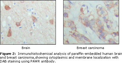
| Cellular Location | Cytoplasm. Nucleus. Note=Mainly cytoplasmic in absence of apoptosis signal and in normal cells. Nuclear in most cancer cell lines. Nuclear entry seems to be essential but not sufficient for apoptosis (By similarity). Nuclear localization includes nucleoplasm and PML nuclear bodies. |
| Tissue Location | Widely expressed. Expression is elevated in various neurodegenerative diseases such as amyotrophic lateral sclerosis, Alzheimer, Parkinson and Huntington diseases and stroke. Down-regulated in several cancers. |

Thousands of laboratories across the world have published research that depended on the performance of antibodies from Abcepta to advance their research. Check out links to articles that cite our products in major peer-reviewed journals, organized by research category.
info@abcepta.com, and receive a free "I Love Antibodies" mug.

Provided below are standard protocols that you may find useful for product applications.
References
1. J Biol Chem. 2004 Jul 2;279(27):28266-75. 2. Exp Hematol. 2004 Jul;32(7):649-56. 3. Mol Cell Biol. 2005 Feb;25(3):1146-61. 4. Psychiatr Genet. 2006 Oct;16(5):193-6.
If you have used an Abcepta product and would like to share how it has performed, please click on the "Submit Review" button and provide the requested information. Our staff will examine and post your review and contact you if needed.
If you have any additional inquiries please email technical services at tech@abcepta.com.
Ordering Information
Other Products
Shipping Information
