


 Foundational characteristics of cancer include proliferation, angiogenesis, migration, evasion of apoptosis, and cellular immortality. Find key markers for these cellular processes and antibodies to detect them.
 The SUMOplot™ Analysis Program predicts and scores sumoylation sites in your protein. SUMOylation is a post-translational modification involved in various cellular processes, such as nuclear-cytosolic transport, transcriptional regulation, apoptosis, protein stability, response to stress, and progression through the cell cycle.
 The Autophagy Receptor Motif Plotter predicts and scores autophagy receptor binding sites in your protein. Identifying proteins connected to this pathway is critical to understanding the role of autophagy in physiological as well as pathological processes such as development, differentiation, neurodegenerative diseases, stress, infection, and cancer.
troponin T2 Antibody
Purified Mouse Monoclonal Antibody
- SPECIFICATION
- CITATIONS
- PROTOCOLS
- BACKGROUND

Application
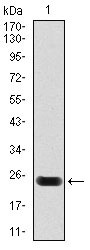
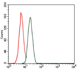
| WB, FC, E |
|---|---|
| Primary Accession | P45379 |
| Reactivity | Human |
| Host | Mouse |
| Clonality | Monoclonal |
| Clone Names | 1G1 |
| Isotype | IgG1 |
| Calculated MW | 36kDa |
| Description | The protein encoded by this gene is the tropomyosin-binding subunit of the troponin complex, which is located on the thin filament of striated muscles and regulates muscle contraction in response to alterations in intracellular calcium ion concentration. Mutations in this gene have been associated with familial hypertrophic cardiomyopathy as well as with dilated cardiomyopathy. Transcripts for this gene undergo alternative splicing that results in many tissue-specific isoforms, however, the full-length nature of some of these variants has not yet been determined. |
| Immunogen | Purified recombinant fragment of human troponin T2 expressed in E. Coli. |
| Formulation | Ascitic fluid containing 0.03% sodium azide. |
| Gene ID | 7139 |
|---|---|
| Other Names | Troponin T, cardiac muscle, TnTc, Cardiac muscle troponin T, cTnT, TNNT2 |
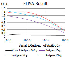
| Dilution | WB~~1/500 - 1/2000 FC~~1/200 - 1/400 E~~1/10000 |
| Storage | Maintain refrigerated at 2-8°C for up to 6 months. For long term storage store at -20°C in small aliquots to prevent freeze-thaw cycles. |
| Precautions | troponin T2 Antibody is for research use only and not for use in diagnostic or therapeutic procedures. |
| Name | TNNT2 |
|---|---|
| Function | Troponin T is the tropomyosin-binding subunit of troponin, the thin filament regulatory complex which confers calcium-sensitivity to striated muscle actomyosin ATPase activity. |
| Tissue Location | Heart. The fetal heart shows a greater expression in the atrium than in the ventricle, while the adult heart shows a greater expression in the ventricle than in the atrium. Isoform 6 predominates in normal adult heart. Isoforms 1, 7 and 8 are expressed in fetal heart. Isoform 7 is also expressed in failing adult heart |

Thousands of laboratories across the world have published research that depended on the performance of antibodies from Abcepta to advance their research. Check out links to articles that cite our products in major peer-reviewed journals, organized by research category.
info@abcepta.com, and receive a free "I Love Antibodies" mug.

Provided below are standard protocols that you may find useful for product applications.
References
Cardiovasc Res. 2010 Jun 1;86(3):452-60. Circ Cardiovasc Genet. 2009 Aug;2(4):306-13.
If you have used an Abcepta product and would like to share how it has performed, please click on the "Submit Review" button and provide the requested information. Our staff will examine and post your review and contact you if needed.
If you have any additional inquiries please email technical services at tech@abcepta.com.
Ordering Information
Other Products
Shipping Information
