


 Foundational characteristics of cancer include proliferation, angiogenesis, migration, evasion of apoptosis, and cellular immortality. Find key markers for these cellular processes and antibodies to detect them.
 The SUMOplot™ Analysis Program predicts and scores sumoylation sites in your protein. SUMOylation is a post-translational modification involved in various cellular processes, such as nuclear-cytosolic transport, transcriptional regulation, apoptosis, protein stability, response to stress, and progression through the cell cycle.
 The Autophagy Receptor Motif Plotter predicts and scores autophagy receptor binding sites in your protein. Identifying proteins connected to this pathway is critical to understanding the role of autophagy in physiological as well as pathological processes such as development, differentiation, neurodegenerative diseases, stress, infection, and cancer.
ENO2 Antibody
Purified Mouse Monoclonal Antibody
- SPECIFICATION
- CITATIONS
- PROTOCOLS
- BACKGROUND

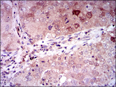
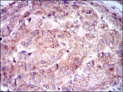
Application
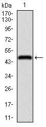
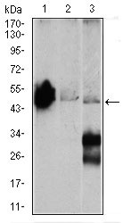
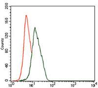
| WB, IHC, FC, E |
|---|---|
| Primary Accession | P09104 |
| Reactivity | Human, Mouse |
| Host | Mouse |
| Clonality | Monoclonal |
| Clone Names | 5D3 |
| Isotype | IgG1 |
| Calculated MW | 47.3kDa |
| Description | This gene encodes one of the three enolase isoenzymes found in mammals. This isoenzyme, a homodimer, is found in mature neurons and cells of neuronal origin. A switch from alpha enolase to gamma enolase occurs in neural tissue during development in rats and primates. |
| Immunogen | Purified recombinant fragment of human ENO2 (AA: 251-433) expressed in E. Coli. |
| Formulation | Purified antibody in PBS with 0.05% sodium azide |
| Gene ID | 2026 |
|---|---|
| Other Names | Gamma-enolase, 4.2.1.11, 2-phospho-D-glycerate hydro-lyase, Enolase 2, Neural enolase, Neuron-specific enolase, NSE, ENO2 |
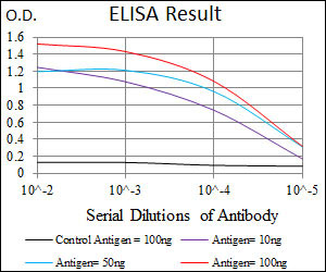
| Dilution | WB~~1/500 - 1/2000 IHC~~1/200 - 1/1000 FC~~1/200 - 1/400 E~~1/10000 |
| Storage | Maintain refrigerated at 2-8°C for up to 6 months. For long term storage store at -20°C in small aliquots to prevent freeze-thaw cycles. |
| Precautions | ENO2 Antibody is for research use only and not for use in diagnostic or therapeutic procedures. |
| Name | ENO2 |
|---|---|
| Function | Has neurotrophic and neuroprotective properties on a broad spectrum of central nervous system (CNS) neurons. Binds, in a calcium- dependent manner, to cultured neocortical neurons and promotes cell survival (By similarity). |
| Cellular Location | Cytoplasm. Cell membrane. Note=Can translocate to the plasma membrane in either the homodimeric (alpha/alpha) or heterodimeric (alpha/gamma) form |
| Tissue Location | The alpha/alpha homodimer is expressed in embryo and in most adult tissues. The alpha/beta heterodimer and the beta/beta homodimer are found in striated muscle, and the alpha/gamma heterodimer and the gamma/gamma homodimer in neurons |

Thousands of laboratories across the world have published research that depended on the performance of antibodies from Abcepta to advance their research. Check out links to articles that cite our products in major peer-reviewed journals, organized by research category.
info@abcepta.com, and receive a free "I Love Antibodies" mug.

Provided below are standard protocols that you may find useful for product applications.
References
1.Neurology. 2011 Aug 16;77(7):623-30. 2.Lung Cancer. 2011 Feb;71(2):224-8.
If you have used an Abcepta product and would like to share how it has performed, please click on the "Submit Review" button and provide the requested information. Our staff will examine and post your review and contact you if needed.
If you have any additional inquiries please email technical services at tech@abcepta.com.
Ordering Information
Other Products
Shipping Information
