


 Foundational characteristics of cancer include proliferation, angiogenesis, migration, evasion of apoptosis, and cellular immortality. Find key markers for these cellular processes and antibodies to detect them.
 The SUMOplot™ Analysis Program predicts and scores sumoylation sites in your protein. SUMOylation is a post-translational modification involved in various cellular processes, such as nuclear-cytosolic transport, transcriptional regulation, apoptosis, protein stability, response to stress, and progression through the cell cycle.
 The Autophagy Receptor Motif Plotter predicts and scores autophagy receptor binding sites in your protein. Identifying proteins connected to this pathway is critical to understanding the role of autophagy in physiological as well as pathological processes such as development, differentiation, neurodegenerative diseases, stress, infection, and cancer.
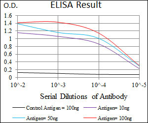
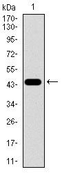
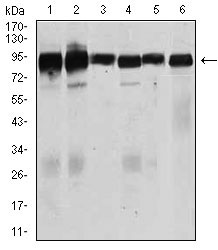
TGFBR3 Antibody
Purified Mouse Monoclonal Antibody
- SPECIFICATION
- CITATIONS
- PROTOCOLS
- BACKGROUND

Application
| WB, E |
|---|---|
| Primary Accession | Q03167 |
| Reactivity | Human, Mouse |
| Host | Mouse |
| Clonality | Monoclonal |
| Clone Names | 1C5H11 |
| Isotype | IgG1 |
| Calculated MW | 93.4kDa |
| Description | This locus encodes the transforming growth factor (TGF)-beta type III receptor. The encoded receptor is a membrane proteoglycan that often functions as a co-receptor with other TGF-beta receptor superfamily members. Ectodomain shedding produces soluble TGFBR3, which may inhibit TGFB signaling. Decreased expression of this receptor has been observed in various cancers. Alternatively spliced transcript variants encoding different isoforms have been identified for this gene. |
| Immunogen | Purified recombinant fragment of human TGFBR3 (AA: 147-328 ) expressed in E. Coli. |
| Formulation | Purified antibody in PBS with 0.05% sodium azide |
| Gene ID | 7049 |
|---|---|
| Other Names | Transforming growth factor beta receptor type 3, TGF-beta receptor type 3, TGFR-3, Betaglycan, Transforming growth factor beta receptor III, TGF-beta receptor type III, TGFBR3 |
| Dilution | WB~~1/500 - 1/2000 E~~1/10000 |
| Storage | Maintain refrigerated at 2-8°C for up to 6 months. For long term storage store at -20°C in small aliquots to prevent freeze-thaw cycles. |
| Precautions | TGFBR3 Antibody is for research use only and not for use in diagnostic or therapeutic procedures. |
| Name | TGFBR3 (HGNC:11774) |
|---|---|
| Function | Cell surface receptor that regulates diverse cellular processes including cell proliferation, differentiation, migration, and apoptosis (PubMed:12958365, PubMed:19416857). Initiates BMP, inhibin, and TGF-beta signaling pathways by interacting with different ligands including TGFB1, BMP2, BMP5, BMP7 or GDF5 (PubMed:18184661). Alternatively, acts as a cell surface coreceptor for BMP ligands, serving to enhance ligand binding by differentially regulating BMPR1A/ALK3 and BMPR1B/ALK6 receptor trafficking (PubMed:19726563). Promotes epithelial cell adhesion, focal adhesion formation and integrin signaling during epithelial cell spreading on fibronectin (PubMed:22562249). By interacting with the scaffolding protein beta- arrestin2/ARRB2, regulates migration or actin cytoskeleton and promotes the activation of CDC42 as well as the inhibition of NF-kappa-B (PubMed:19416857, PubMed:19325136). In gonadotrope cells, acts as an inhibin A coreceptor and regulates follicle-stimulating hormone (FSH) levels and female fertility (By similarity). Plays a role in the inhibition of directed and random cell migration in epithelial cells by altering the actin cytoskeletal organization (PubMed:19416857). Participates in epithelial-mesenchymal transformation (EMT) upon binding to BMP2 or TGFB2, by activating the PAR6/SMURF1/RHOA pathway (By similarity). |
| Cellular Location | Cell membrane; Single-pass type I membrane protein. Secreted {ECO:0000250|UniProtKB:P26342}. Secreted, extracellular space, extracellular matrix {ECO:0000250|UniProtKB:P26342}. Note=Exists both as a membrane-bound form and as soluble form in serum and in the extracellular matrix. {ECO:0000250|UniProtKB:P26342} |

Thousands of laboratories across the world have published research that depended on the performance of antibodies from Abcepta to advance their research. Check out links to articles that cite our products in major peer-reviewed journals, organized by research category.
info@abcepta.com, and receive a free "I Love Antibodies" mug.

Provided below are standard protocols that you may find useful for product applications.
References
1.Mol Biol Cell. 2011 May;22(9):1463-72. 2.Zhongguo Fei Ai Za Zhi. 2010 May;13(5):451-7.
If you have used an Abcepta product and would like to share how it has performed, please click on the "Submit Review" button and provide the requested information. Our staff will examine and post your review and contact you if needed.
If you have any additional inquiries please email technical services at tech@abcepta.com.
Ordering Information
Other Products
Shipping Information
