


 Foundational characteristics of cancer include proliferation, angiogenesis, migration, evasion of apoptosis, and cellular immortality. Find key markers for these cellular processes and antibodies to detect them.
 The SUMOplot™ Analysis Program predicts and scores sumoylation sites in your protein. SUMOylation is a post-translational modification involved in various cellular processes, such as nuclear-cytosolic transport, transcriptional regulation, apoptosis, protein stability, response to stress, and progression through the cell cycle.
 The Autophagy Receptor Motif Plotter predicts and scores autophagy receptor binding sites in your protein. Identifying proteins connected to this pathway is critical to understanding the role of autophagy in physiological as well as pathological processes such as development, differentiation, neurodegenerative diseases, stress, infection, and cancer.
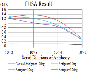
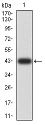
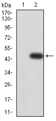
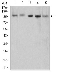
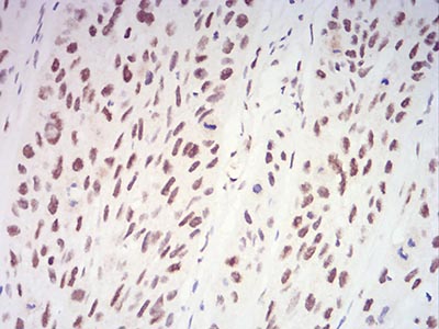
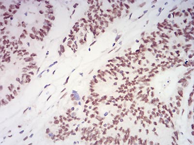
Mouse Monoclonal Antibody to XRN2
Purified Mouse Monoclonal Antibody
- SPECIFICATION
- CITATIONS
- PROTOCOLS
- BACKGROUND

Application
| WB, IHC, E |
|---|---|
| Primary Accession | Q9H0D6 |
| Reactivity | Human |
| Host | Mouse |
| Clonality | Monoclonal |
| Clone Names | 9F7G11 |
| Isotype | Mouse IgG1 |
| Calculated MW | 108.5kDa |
| Description | This gene encodes a 5'-3' exonuclease that promotes transcription termination at cotranscriptional cleavage sites. Alternative splicing results in multiple transcript variants encoding different isoforms.; |
| Immunogen | Purified recombinant fragment of human XRN2 (AA: 398-547) expressed in E. Coli. |
| Formulation | Purified antibody in PBS with 0.05% sodium azide |
| Application Note | ELISA: 1/10000 WB: 1/500 - 1/2000 IHC: 1/200 - 1/1000 |
| Gene ID | 22803 |
|---|---|
| Dilution | WB~~1:1000 IHC~~1:100~500 E~~N/A |
| Storage | Maintain refrigerated at 2-8°C for up to 6 months. For long term storage store at -20°C in small aliquots to prevent freeze-thaw cycles. |
| Precautions | Mouse Monoclonal Antibody to XRN2 is for research use only and not for use in diagnostic or therapeutic procedures. |
| Name | XRN2 |
|---|---|
| Function | Possesses 5'->3' exoribonuclease activity (By similarity). May promote the termination of transcription by RNA polymerase II. During transcription termination, cleavage at the polyadenylation site liberates a 5' fragment which is subsequently processed to form the mature mRNA and a 3' fragment which remains attached to the elongating polymerase. The processive degradation of this 3' fragment by this protein may promote termination of transcription. Binds to RNA polymerase II (RNAp II) transcription termination R-loops formed by G- rich pause sites (PubMed:21700224). |
| Cellular Location | Nucleus, nucleolus. |
| Tissue Location | Expressed in the spleen, thymus, prostate, testis, ovary, small intestine, colon, peripheral blood leukocytes, heart, brain, placenta, lung, liver, skeletal muscle, kidney, and pancreas Isoform 2 is expressed predominantly in peripheral blood leukocytes |

Thousands of laboratories across the world have published research that depended on the performance of antibodies from Abcepta to advance their research. Check out links to articles that cite our products in major peer-reviewed journals, organized by research category.
info@abcepta.com, and receive a free "I Love Antibodies" mug.

Provided below are standard protocols that you may find useful for product applications.
References
1.EMBO J. 2012 May 30;31(11):2566-78. ; 2.DNA Seq. 2005 Apr;16(2):143-6.;
If you have used an Abcepta product and would like to share how it has performed, please click on the "Submit Review" button and provide the requested information. Our staff will examine and post your review and contact you if needed.
If you have any additional inquiries please email technical services at tech@abcepta.com.
Ordering Information
Other Products
Shipping Information
